Angelova Polina, Kehayov Ivo, Ordonez-Rubiano Edgar G, Figueredo Luisa F, Zlatareva Dora
Department of Neurosurgery, Faculty of Medicine, Medical University of Plovdiv, Plovdiv, Bulgaria.
Department of Neurosurgery, Hospital de San José - Sociedad de Cirugía de Bogotá, Fundación Universitaria de Ciencias de la Salud, Bogotá D.C., Colombia.
Korean J Neurotrauma. 2023 Jun 22;19(2):249-257. doi: 10.13004/kjnt.2023.19.e27. eCollection 2023 Jun.
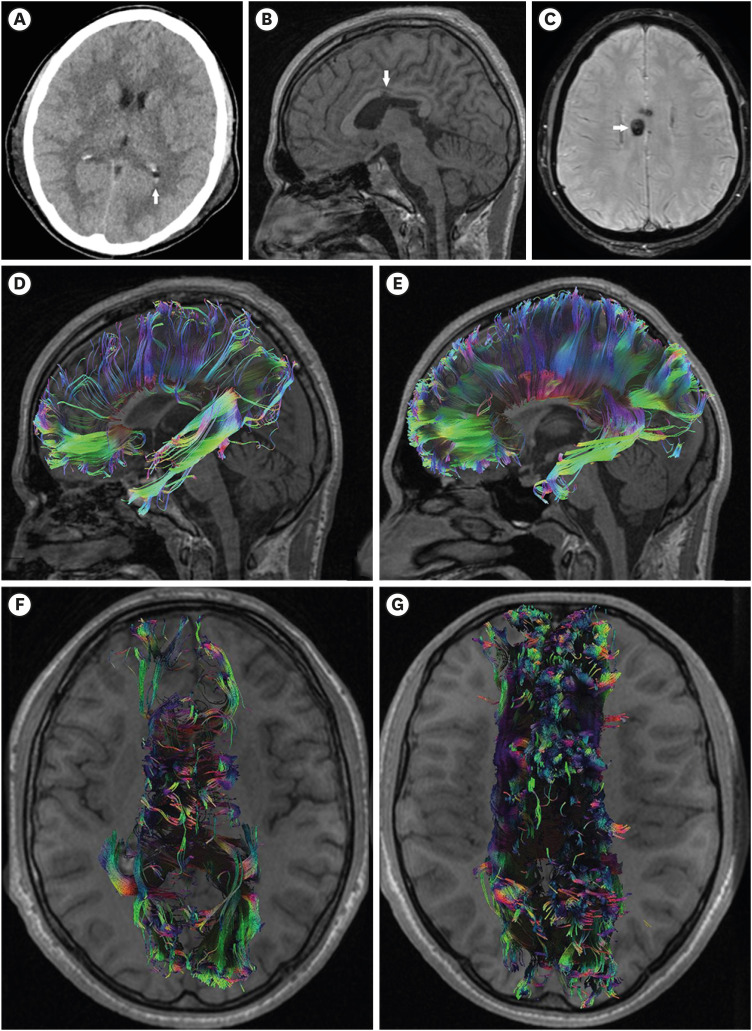
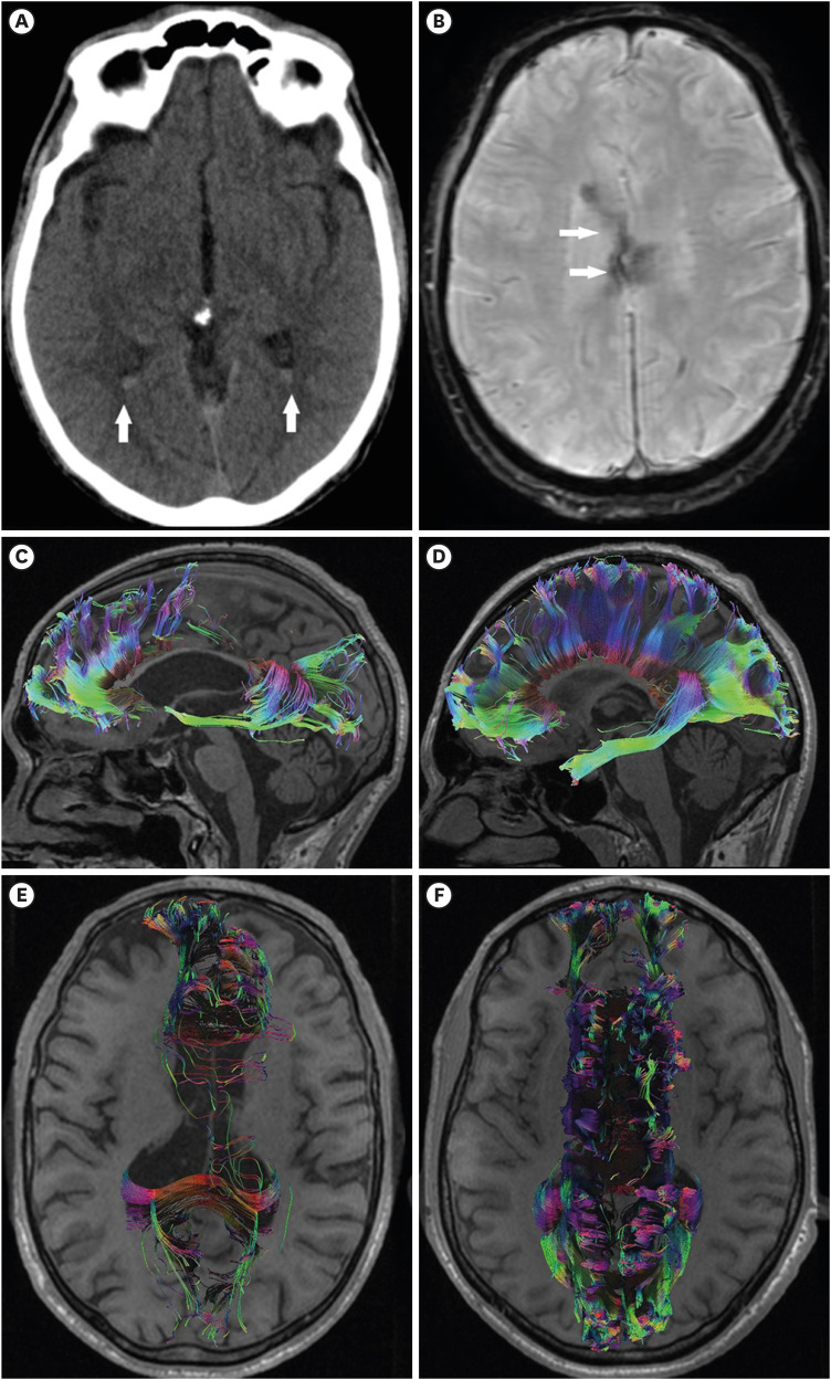
Severe traumatic brain injury (TBI) is often associated with diffuse axonal injury. Diffuse axonal injury affecting the corpus callosum may present with intraventricular hemorrhage on baseline computed tomography (CT) scan. Posttraumatic corpus callosum damage is a chronic condition that can be diagnosed over the long term using various magnetic resonance imaging (MRI) sequences. Here, we present two cases of severe survivors of TBI with isolated intraventricular hemorrhage detected on an initial CT scan. After acute trauma management, long-term follow-up was performed. Diffusion tensor imaging and subsequent tractography revealed a significant decrease in the fractional anisotropy values and the number of corpus callosum fibers compared with those in healthy control patients. This study presents a possible correlation between traumatic intraventricular hemorrhage on admission CT and long-term corpus callosum impairment detected on MRI in patients with severe head injury by presenting demonstrative cases and conducting a literature review.
严重创伤性脑损伤(TBI)常与弥漫性轴索损伤相关。影响胼胝体的弥漫性轴索损伤在基线计算机断层扫描(CT)上可能表现为脑室内出血。创伤后胼胝体损伤是一种慢性疾病,可通过多种磁共振成像(MRI)序列进行长期诊断。在此,我们报告两例严重TBI幸存者,他们在初次CT扫描时发现孤立性脑室内出血。在进行急性创伤处理后,进行了长期随访。弥散张量成像及随后的纤维束成像显示,与健康对照患者相比,分数各向异性值和胼胝体纤维数量显著减少。本研究通过展示实例并进行文献综述,提出了重度颅脑损伤患者入院时CT上的创伤性脑室内出血与MRI上检测到的长期胼胝体损伤之间可能存在的相关性。