• 文献检索
  • 文档翻译
  • 深度研究
  • 学术资讯
  • Suppr Zotero 插件Zotero 插件
  • 邀请有礼
  • 套餐&价格
  • 历史记录
应用&插件
Suppr Zotero 插件Zotero 插件浏览器插件Mac 客户端Windows 客户端微信小程序
定价
高级版会员购买积分包购买API积分包
服务
文献检索文档翻译深度研究API 文档MCP 服务
关于我们
关于 Suppr公司介绍联系我们用户协议隐私条款
关注我们

Suppr 超能文献

核心技术专利:CN118964589B侵权必究
粤ICP备2023148730 号-1Suppr @ 2026

文献检索

告别复杂PubMed语法,用中文像聊天一样搜索,搜遍4000万医学文献。AI智能推荐,让科研检索更轻松。

立即免费搜索

文件翻译

保留排版,准确专业,支持PDF/Word/PPT等文件格式,支持 12+语言互译。

免费翻译文档

深度研究

AI帮你快速写综述,25分钟生成高质量综述,智能提取关键信息,辅助科研写作。

立即免费体验

淋巴水肿大鼠模型的建立及基于胶原蛋白的医疗器械植入用于治疗干预

Development of a rat model of lymphedema and the implantation of a collagen-based medical device for therapeutic intervention.

作者信息

Nguyen Dung, Dionyssiou Dimitrios, Zaitseva Tatiana S, Zhou Anna T, Sue Gloria, Deptula Peter, Moroz Maxim A, Tabada Peter, Rockson Stanley G, Paukshto Michael V, Cheng Ming-Huei, Huang Ngan F

机构信息

Department of Plastic and Reconstructive Surgery, Stanford University, Stanford, CA, United States.

Department of Plastic Surgery, Aristotle University of Thessaloniki, Greece.

出版信息

Front Cardiovasc Med. 2023 Jul 4;10:1214116. doi: 10.3389/fcvm.2023.1214116. eCollection 2023.

DOI:10.3389/fcvm.2023.1214116
PMID:37469481
原文链接:https://pmc.ncbi.nlm.nih.gov/articles/PMC10353614/
Abstract

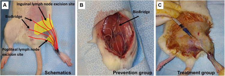
Secondary lymphedema is a common condition among cancer survivors, and treatment strategies to prevent or treat lymphedema are in high demand. The development of novel strategies to diagnose or treat lymphedema would benefit from a robust experimental animal model of secondary lymphedema. The purpose of this methods paper is to describe and summarize our experience in developing and characterizing a rat hindlimb model of lymphedema. Here we describe a protocol to induce secondary lymphedema that takes advantage of micro computed tomography imaging for limb volume measurements and visualization of lymph drainage with near infrared imaging. To demonstrate the utility of this preclinical model for studying the therapeutic benefit of novel devices, we apply this animal model to test the efficacy of a biomaterials-based implantable medical device.

摘要

继发性淋巴水肿是癌症幸存者中的常见病症,因此对预防或治疗淋巴水肿的治疗策略有很高的需求。开发用于诊断或治疗淋巴水肿的新策略将受益于强大的继发性淋巴水肿实验动物模型。本方法论文的目的是描述和总结我们在开发和表征淋巴水肿大鼠后肢模型方面的经验。在这里,我们描述了一种诱导继发性淋巴水肿的方案,该方案利用微型计算机断层扫描成像进行肢体体积测量,并通过近红外成像可视化淋巴引流。为了证明这种临床前模型在研究新型设备治疗益处方面的实用性,我们应用此动物模型来测试一种基于生物材料的可植入医疗设备的疗效。

https://cdn.ncbi.nlm.nih.gov/pmc/blobs/5b88/10353614/f8346adcf4f3/fcvm-10-1214116-g005.jpg
https://cdn.ncbi.nlm.nih.gov/pmc/blobs/5b88/10353614/b454f4ae7b73/fcvm-10-1214116-g001.jpg
https://cdn.ncbi.nlm.nih.gov/pmc/blobs/5b88/10353614/96536fa2d862/fcvm-10-1214116-g002.jpg
https://cdn.ncbi.nlm.nih.gov/pmc/blobs/5b88/10353614/bcc2a48e7ea5/fcvm-10-1214116-g003.jpg
https://cdn.ncbi.nlm.nih.gov/pmc/blobs/5b88/10353614/997d8738c150/fcvm-10-1214116-g004.jpg
https://cdn.ncbi.nlm.nih.gov/pmc/blobs/5b88/10353614/f8346adcf4f3/fcvm-10-1214116-g005.jpg

相似文献

1
Development of a rat model of lymphedema and the implantation of a collagen-based medical device for therapeutic intervention.淋巴水肿大鼠模型的建立及基于胶原蛋白的医疗器械植入用于治疗干预
Front Cardiovasc Med. 2023 Jul 4;10:1214116. doi: 10.3389/fcvm.2023.1214116. eCollection 2023.
2
Lymphatic regeneration after implantation of aligned nanofibrillar collagen scaffolds: Preliminary preclinical and clinical results.植入取向纳米纤维胶原支架后的淋巴再生:初步的临床前和临床结果。
J Surg Oncol. 2022 Feb;125(2):113-122. doi: 10.1002/jso.26679. Epub 2021 Sep 21.
3
Treatment of Rat Lymphedema by Propeller Lymphatic Tissue Flap Combined with Nanofibrillar Collagen Scaffolds.螺旋桨式淋巴组织瓣联合纳米纤维胶原支架治疗大鼠淋巴水肿
J Reconstr Microsurg. 2024 Feb;40(2):145-155. doi: 10.1055/a-2086-0269. Epub 2023 May 4.
4
Experimental Drainage Device to Reduce Lymphoedema in a Rat Model.实验性引流装置减少大鼠模型中的淋巴水肿。
Eur J Vasc Endovasc Surg. 2019 Jun;57(6):859-867. doi: 10.1016/j.ejvs.2018.04.014. Epub 2018 May 25.
5
Aligned nanofibrillar collagen scaffolds - Guiding lymphangiogenesis for treatment of acquired lymphedema.排列的纳米纤维胶原支架——引导淋巴管生成以治疗后天性淋巴水肿
Biomaterials. 2016 Sep;102:259-267. doi: 10.1016/j.biomaterials.2016.05.040. Epub 2016 Jun 7.
6
Enhancing lymphangiogenesis and lymphatic drainage to vascularized lymph nodes with nanofibrillar collagen scaffolds.利用纳米纤维胶原支架增强血管化淋巴结的淋巴管生成和淋巴引流。
J Surg Oncol. 2022 Dec;126(7):1169-1175. doi: 10.1002/jso.27058. Epub 2022 Aug 11.
7
Popliteal Vascular Lymph Node Resection in the Rabbit Hindlimb for Secondary Lymphedema Induction.兔后肢用于继发性淋巴水肿诱导的腘血管淋巴结切除术。
J Vis Exp. 2022 Nov 30(189). doi: 10.3791/64576.
8
Refinement of a rodent model of peripheral lymphedema.外周性淋巴水肿啮齿动物模型的优化
Lymphology. 1999 Sep;32(3):111-7.
9
Nanofibrillar Collagen Scaffold Enhances Edema Reduction and Formation of New Lymphatic Collectors after Lymphedema Surgery.纳米纤维胶原支架可减少淋巴水肿手术后的水肿,并促进新淋巴管生成。
Plast Reconstr Surg. 2021 Dec 1;148(6):1382-1393. doi: 10.1097/PRS.0000000000008590.
10
Quantification of Chronic Lymphedema in a Revised Mouse Model.改良小鼠模型中慢性淋巴水肿的定量分析
Ann Plast Surg. 2018 Nov;81(5):594-603. doi: 10.1097/SAP.0000000000001537.

引用本文的文献

1
Lymphatic Regeneration after Popliteal Lymph Node Excision and Implantation of Aligned Nanofibrillar Collagen Scaffolds: An Experimental Rabbit Model.腘淋巴结切除及植入排列整齐的纳米纤维胶原支架后的淋巴再生:实验性兔模型
J Funct Biomater. 2024 Aug 21;15(8):235. doi: 10.3390/jfb15080235.
2
Editorial for the Special Issue "Vasorum Lymphaticorum: From the Discovery of the Lymphatic System to the New Perspectives in Microsurgical Reconstruction and Patient Rehabilitation".专刊编辑寄语:从淋巴管系统的发现到显微重建和患者康复的新视角。
Medicina (Kaunas). 2024 Feb 10;60(2):307. doi: 10.3390/medicina60020307.

本文引用的文献

1
Lymphatic channel sheet of polydimethylsiloxane for preventing secondary lymphedema in the rat upper limb model.用于预防大鼠上肢模型继发性淋巴水肿的聚二甲基硅氧烷淋巴通道片
Bioeng Transl Med. 2022 Jul 5;8(1):e10371. doi: 10.1002/btm2.10371. eCollection 2023 Jan.
2
Popliteal Vascular Lymph Node Resection in the Rabbit Hindlimb for Secondary Lymphedema Induction.兔后肢用于继发性淋巴水肿诱导的腘血管淋巴结切除术。
J Vis Exp. 2022 Nov 30(189). doi: 10.3791/64576.
3
Networked lymphatic endothelial cells in a transplanted cell sheet contribute to form functional lymphatic vessels.
网络状的淋巴管内皮细胞在移植的细胞片中有助于形成功能性的淋巴管。
Sci Rep. 2022 Dec 15;12(1):21698. doi: 10.1038/s41598-022-26041-0.
4
Single and combined impacts of irradiation and surgery on lymphatic vasculature and fibrosis associated to secondary lymphedema.辐射与手术对与继发性淋巴水肿相关的淋巴管系统和纤维化的单一及联合影响。
Front Pharmacol. 2022 Oct 18;13:1016138. doi: 10.3389/fphar.2022.1016138. eCollection 2022.
5
Enhancing lymphangiogenesis and lymphatic drainage to vascularized lymph nodes with nanofibrillar collagen scaffolds.利用纳米纤维胶原支架增强血管化淋巴结的淋巴管生成和淋巴引流。
J Surg Oncol. 2022 Dec;126(7):1169-1175. doi: 10.1002/jso.27058. Epub 2022 Aug 11.
6
Nanofibrillar Collagen Scaffold Enhances Edema Reduction and Formation of New Lymphatic Collectors after Lymphedema Surgery.纳米纤维胶原支架可减少淋巴水肿手术后的水肿,并促进新淋巴管生成。
Plast Reconstr Surg. 2021 Dec 1;148(6):1382-1393. doi: 10.1097/PRS.0000000000008590.
7
Lymphatic regeneration after implantation of aligned nanofibrillar collagen scaffolds: Preliminary preclinical and clinical results.植入取向纳米纤维胶原支架后的淋巴再生:初步的临床前和临床结果。
J Surg Oncol. 2022 Feb;125(2):113-122. doi: 10.1002/jso.26679. Epub 2021 Sep 21.
8
Tissue Engineering Strategies for Cancer-Related Lymphedema.癌症相关淋巴水肿的组织工程策略。
Tissue Eng Part A. 2021 Apr;27(7-8):489-499. doi: 10.1089/ten.TEA.2020.0378.
9
Tissue Engineering of Lymphatic Vasculature in the Arteriovenous Loop Model of the Rat.大鼠动静脉环模型中淋巴管系统的组织工程
Tissue Eng Part A. 2021 Jan;27(1-2):129-141. doi: 10.1089/ten.TEA.2020.0108. Epub 2020 Sep 21.
10
The role of adjunct nanofibrillar collagen scaffold implantation in the surgical management of secondary lymphedema: Review of the literature and summary of initial pilot studies.辅助纳米纤维胶原支架植入在继发性淋巴水肿手术治疗中的作用:文献回顾和初步试点研究总结。
J Surg Oncol. 2020 Jan;121(1):121-128. doi: 10.1002/jso.25576. Epub 2019 Jun 18.