Department of Radiology, Charité - Universitätsmedizin Berlin, Campus Mitte, Humboldt-Universität Zu Berlin, Freie Universität Berlin, 10117, Berlin, Germany.
Canon Medical Systems, Europe BV, Zoetermeer, Netherlands.
Eur Radiol Exp. 2023 Jul 24;7(1):43. doi: 10.1186/s41747-023-00348-7.
To investigate the influence of iodinated contrast medium (ICM) on detection of monosodium urate (MSU) with dual-energy computed tomography (DECT) in two types of phantoms and demonstrate an example patient for clinical illustration.
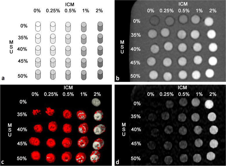
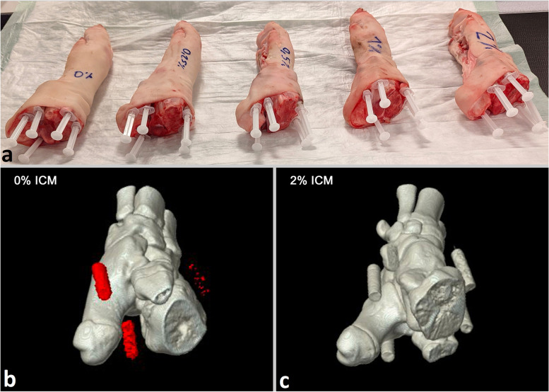
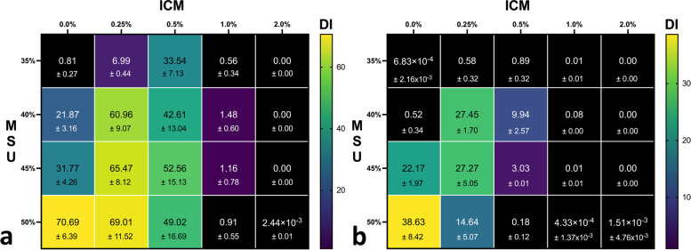
Approval is by the institutional review board, and written informed consent was obtained. A grid-like and a biophantom with 25 suspensions containing different concentrations of ICM (0 to 2%) and MSU (0 to 50%) were prepared and scanned with sequential single-source DECT using established methodology. Ascending orders of tube currents were applied at 80 kVp (16.5 to 220.0 mAs) and 135 kVp (2.75 to 19.25 mAs). Volume and mass measurements were performed using clinical gout software (dual-energy decomposition analysis). Numbers of true-positive and false-positive MSU detections were recorded and compared for different ICM concentrations. We demonstrate a patient with gouty arthritis for clinical illustration.
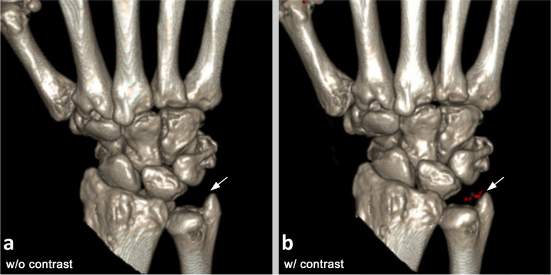
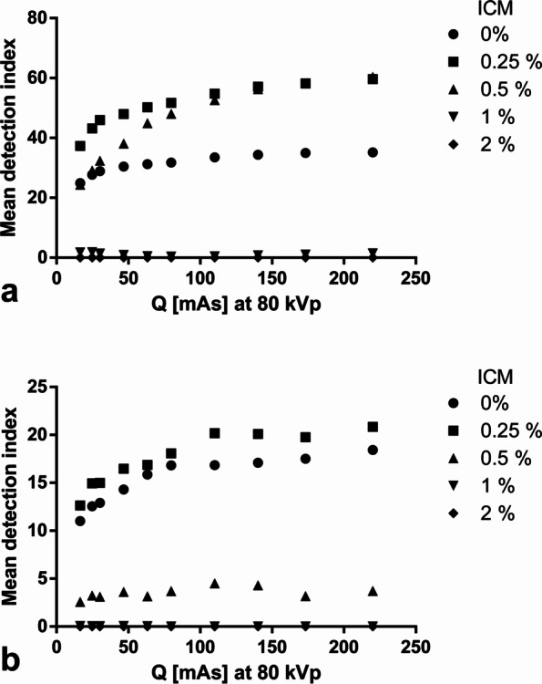
Effects of ICM on MSU detection varied with the amount of iodine. Lower ICM concentrations (0.25 and 0.50%) improved detection of small uric acid concentrations of 35 to 45% in comparison to scans without ICM. However, high ICM concentrations (1 and 2%) almost completely precluded MSU detection for all MSU concentrations investigated. In a patient with gouty arthritis, tophi in the wrist were only detected after intravenous ICM administration.
Exploring multimodal DECT for arthritis imaging, enhancement of ICM influences tophus detection. It can help in visualizing previously undetected MSU depositions but, with too strong enhancement, also obscure tophi.
Use of iodinated contrast media in dual-energy CT might help in visualizing previously undetected uric acid depositions but, with too strong enhancement, obscure gouty tophi.
• Iodine significantly influences the uric acid crystal detection in systematic phantom studies. • Lower iodine concentrations improved detection of low and medium uric acid concentrations. • High concentrations of iodine hampered detection of all uric acid concentrations.
本研究旨在通过双能 CT(DECT)探讨两种模型中碘造影剂(ICM)对单钠尿酸盐(MSU)检测的影响,并通过临床实例加以说明。
本研究获得机构审查委员会批准,并获得书面知情同意。制备了网格状和生物模型,每个模型内均包含 25 个不同浓度的 ICM(0 至 2%)和 MSU(0 至 50%)混悬液,并采用既定方法进行序贯单源 DECT 扫描。管电流递增顺序为 80 kVp(16.5 至 220.0 mAs)和 135 kVp(2.75 至 19.25 mAs)。使用临床痛风软件(双能量分解分析)进行体积和质量测量。记录并比较不同 ICM 浓度下的真阳性和假阳性 MSU 检测结果。我们展示了一位痛风关节炎患者的临床实例。
ICM 对 MSU 检测的影响因碘含量而异。与不使用 ICM 的扫描相比,低浓度的 ICM(0.25% 和 0.50%)可提高 35%至 45%的小尿酸浓度的检测率。然而,高浓度的 ICM(1%和 2%)几乎完全排除了所有研究 MSU 浓度的 MSU 检测。在一位痛风关节炎患者中,腕关节痛风石仅在静脉注射 ICM 后才被检测到。
探索多模态 DECT 进行关节炎成像时,增强 ICM 会影响痛风石的检测。它可以帮助可视化先前未检测到的 MSU 沉积,但增强过度也会使痛风石模糊。
在双能 CT 中使用碘造影剂可能有助于可视化先前未检测到的尿酸沉积,但增强过度会使痛风石模糊。
在系统的体模研究中,碘对尿酸晶体检测有显著影响。
较低的碘浓度可提高低浓度和中浓度尿酸的检测率。
高浓度的碘会阻碍所有尿酸浓度的检测。