Department of Orthopaedics, Zigong Fourth People's Hospital, Zigong, 643000, Sichuan Province, People's Republic of China.
Department of Orthopedics, The Affiliated Traditional Chinese Medicine Hospital of Southwest Medical University, No. 182, Chunhui Road, Luzhou, Sichuan Province, 646000, People's Republic of China.
BMC Musculoskelet Disord. 2023 Jul 24;24(1):602. doi: 10.1186/s12891-023-06615-3.
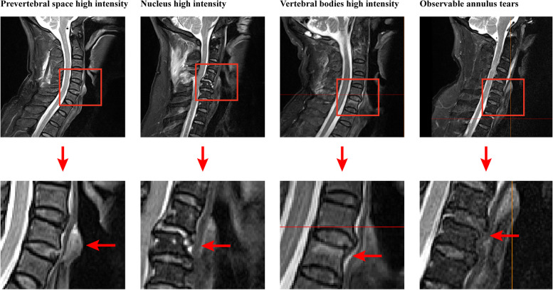
Segmental fusion operations assume paramount significance for individuals afflicted by full layers of annulus tears as they avert the perils of rapid disc degeneration and segmental instability. Structures with high signal intensity in the T2-weighted MRI can predict potential damage to the injured segment. Since local structures are shortly related biomechanically, this may be an effective predictor for annulus tears.
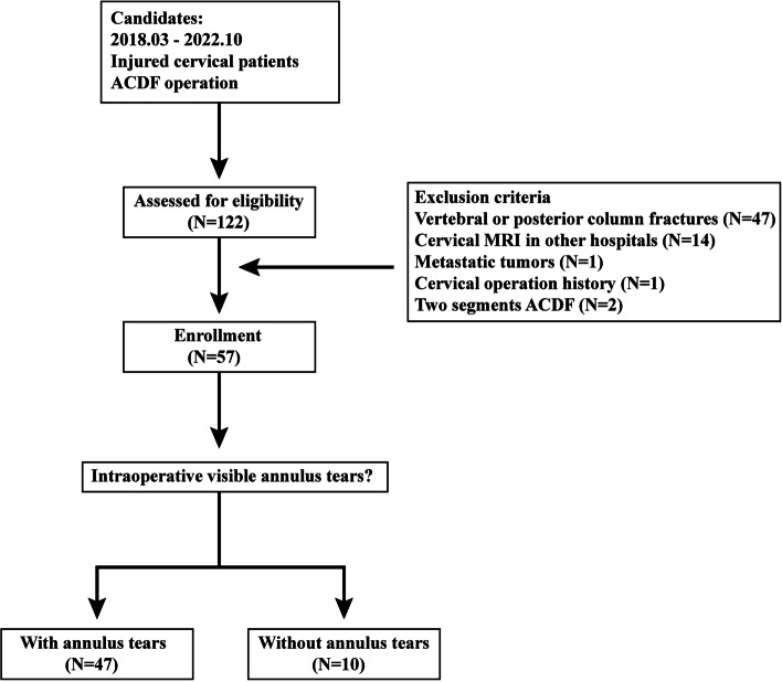
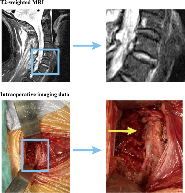
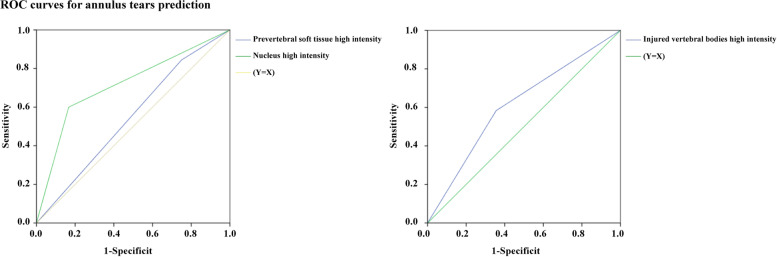
A retrospective analysis of the clinical data of 57 patients afflicted by cervical injuries and subjected to single-segment ACDF has been performed in this study. The surgeon performed intraoperative exploration to assess the integration status of the annulus. The signal intensity of the prevertebral space, nucleus, and injured vertebral bodies were judged in the T2-weighted imaging data. Regression analyses identified independent predictors for annulus tears, and the area under the receiver operating characteristic curve (AUC) was computed to evaluate the predictive performance of potential independent predictors.
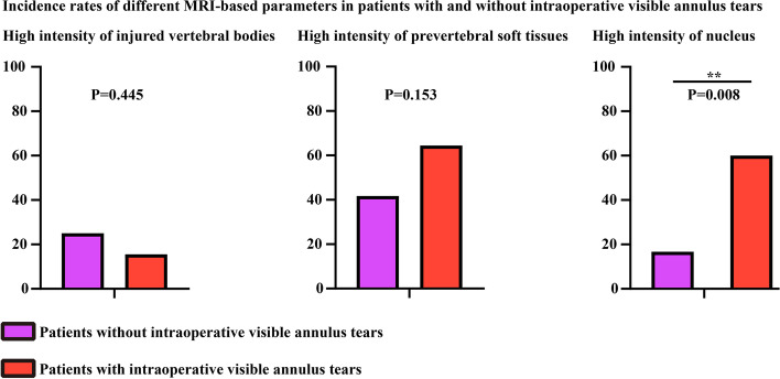
The occurrence of nucleus high intensity was significantly higher among individuals with annulus tears, and the nucleus high intensity was deemed an independent predictor for determining the presence of intraoperative visible annulus tears in patients with cervical injuries. AUC for nucleus high intensity was calculated as 0.717, with a corresponding p-value less than 0.05.
In the realm of diagnosing annulus tears in injured cervical patients, nucleus high intensity in the T2-weighted MRI emerges as a promising predictive factor. Notably, this applies specifically to patients devoid of fracture and visible annulus tears in their MRI scans. Such positive outcomes should be regarded as prospective indications for ACDF.
对于全层纤维环撕裂的患者,节段融合手术至关重要,因为它可以避免椎间盘快速退变和节段不稳定的风险。T2 加权 MRI 中高信号强度的结构可以预测损伤节段的潜在损伤。由于局部结构在生物力学上密切相关,因此这可能是纤维环撕裂的有效预测指标。
本研究对 57 例颈损伤行单节段 ACDF 的患者的临床资料进行了回顾性分析。手术医生在术中探查评估纤维环的融合状态。根据 T2 加权成像数据判断椎前间隙、核和损伤椎体的信号强度。回归分析确定了纤维环撕裂的独立预测因素,并计算了受试者工作特征曲线下的面积(AUC),以评估潜在独立预测因素的预测性能。
纤维环撕裂患者的核高信号发生率明显更高,核高信号是判断颈损伤患者术中可见纤维环撕裂的独立预测因素。核高信号的 AUC 为 0.717,相应的 p 值小于 0.05。
在诊断颈损伤患者的纤维环撕裂方面,T2 加权 MRI 中的核高信号是一个有前途的预测因素。值得注意的是,这特别适用于 MRI 扫描无骨折和可见纤维环撕裂的患者。这些阳性结果应被视为 ACDF 的前瞻性指征。