• 文献检索
  • 文档翻译
  • 深度研究
  • 学术资讯
  • Suppr Zotero 插件Zotero 插件
  • 邀请有礼
  • 套餐&价格
  • 历史记录
应用&插件
Suppr Zotero 插件Zotero 插件浏览器插件Mac 客户端Windows 客户端微信小程序
定价
高级版会员购买积分包购买API积分包
服务
文献检索文档翻译深度研究API 文档MCP 服务
关于我们
关于 Suppr公司介绍联系我们用户协议隐私条款
关注我们

Suppr 超能文献

核心技术专利:CN118964589B侵权必究
粤ICP备2023148730 号-1Suppr @ 2026

文献检索

告别复杂PubMed语法,用中文像聊天一样搜索,搜遍4000万医学文献。AI智能推荐,让科研检索更轻松。

立即免费搜索

文件翻译

保留排版,准确专业,支持PDF/Word/PPT等文件格式,支持 12+语言互译。

免费翻译文档

深度研究

AI帮你快速写综述,25分钟生成高质量综述,智能提取关键信息,辅助科研写作。

立即免费体验

比较单细胞分析揭示了独特的心脏驻留巨噬细胞,对斑马鱼心脏再生至关重要。

Comparative single-cell profiling reveals distinct cardiac resident macrophages essential for zebrafish heart regeneration.

机构信息

Graduate Institute of Life Sciences, National Defense Medical Center, Taipei, Taiwan.

Institute of Biomedical Sciences, Academia Sinica, Taipei, Taiwan.

出版信息

Elife. 2023 Jul 27;12:e84679. doi: 10.7554/eLife.84679.

DOI:10.7554/eLife.84679
PMID:37498060
原文链接:https://pmc.ncbi.nlm.nih.gov/articles/PMC10411971/
Abstract

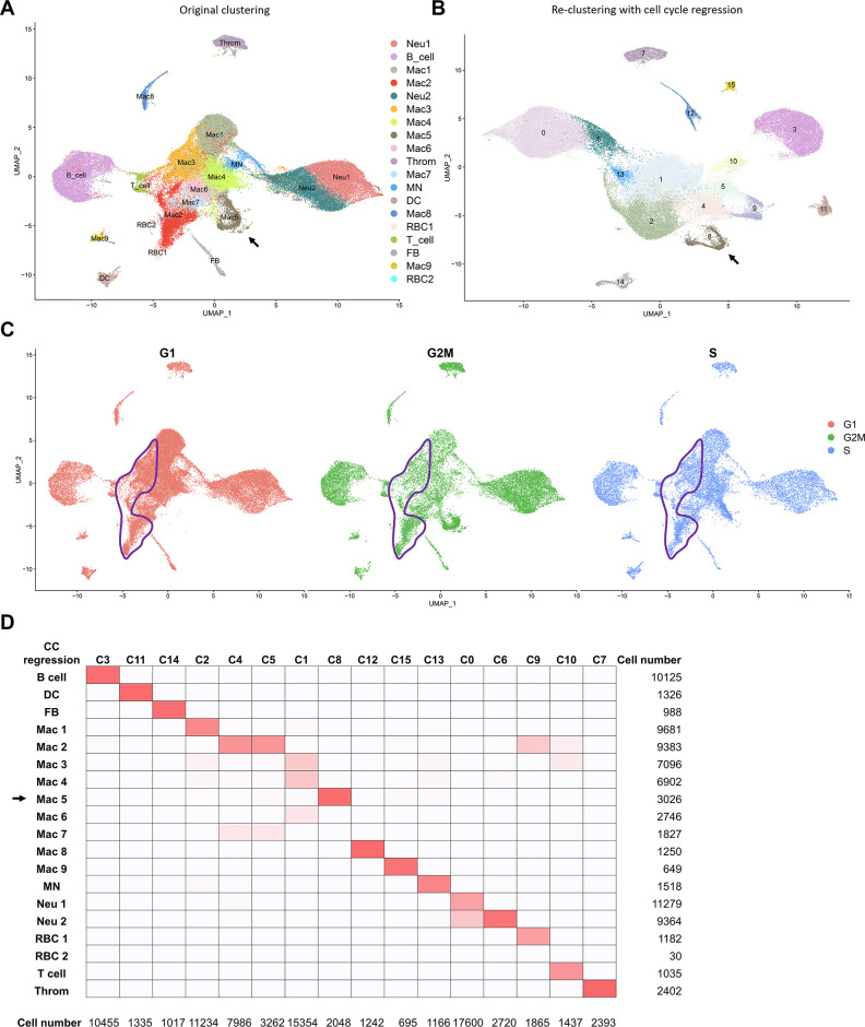
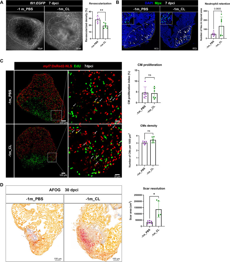
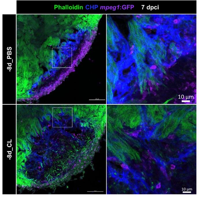
Zebrafish exhibit a robust ability to regenerate their hearts following injury, and the immune system plays a key role in this process. We previously showed that delaying macrophage recruitment by clodronate liposome (-1d_CL, macrophage-delayed model) impairs neutrophil resolution and heart regeneration, even when the infiltrating macrophage number was restored within the first week post injury (Lai et al., 2017). It is thus intriguing to learn the regenerative macrophage property by comparing these late macrophages vs. control macrophages during cardiac repair. Here, we further investigate the mechanistic insights of heart regeneration by comparing the non-regenerative macrophage-delayed model with regenerative controls. Temporal RNAseq analyses revealed that -1d_CL treatment led to disrupted inflammatory resolution, reactive oxygen species homeostasis, and energy metabolism during cardiac repair. Comparative single-cell RNAseq profiling of inflammatory cells from regenerative vs. non-regenerative hearts further identified heterogeneous macrophages and neutrophils, showing alternative activation and cellular crosstalk leading to neutrophil retention and chronic inflammation. Among macrophages, two residential subpopulations ( Mac and Mac 3) were enriched only in regenerative hearts and barely recovered after +1d_CL treatment. To deplete the resident macrophage without delaying the circulating macrophage recruitment, we established the resident macrophage-deficient model by administrating CL earlier at 8 d (-8d_CL) before cryoinjury. Strikingly, resident macrophage-deficient zebrafish still exhibited defects in revascularization, cardiomyocyte survival, debris clearance, and extracellular matrix remodeling/scar resolution without functional compensation from the circulating/monocyte-derived macrophages. Our results characterized the diverse function and interaction between inflammatory cells and identified unique resident macrophages prerequisite for zebrafish heart regeneration.

摘要

斑马鱼在受伤后表现出强大的心脏再生能力,而免疫系统在这个过程中起着关键作用。我们之前曾表明,通过使用氯膦酸盐脂质体(-1d_CL,巨噬细胞延迟模型)延迟巨噬细胞募集会损害中性粒细胞的清除和心脏再生,即使在损伤后第一周内恢复了浸润巨噬细胞数量(Lai 等人,2017)。因此,通过比较心脏修复过程中的这些晚期巨噬细胞与对照巨噬细胞,了解再生巨噬细胞的特性是很有趣的。在这里,我们通过比较非再生性巨噬细胞延迟模型与再生性对照模型,进一步研究心脏再生的机制见解。时间 RNAseq 分析表明,-1d_CL 处理导致心脏修复过程中炎症反应的清除、活性氧稳态和能量代谢受到破坏。对来自再生性和非再生性心脏的炎症细胞进行比较单细胞 RNAseq 分析进一步鉴定了异质性巨噬细胞和中性粒细胞,显示出替代性激活和细胞串扰,导致中性粒细胞滞留和慢性炎症。在巨噬细胞中,仅在再生性心脏中富集了两个常驻亚群(Mac 和 Mac3),并且在 +1d_CL 处理后几乎没有恢复。为了在不延迟循环巨噬细胞募集的情况下耗尽常驻巨噬细胞,我们通过在冷冻损伤前 8 天(-8d_CL)更早地给予 CL 建立了常驻巨噬细胞缺陷模型。令人惊讶的是,常驻巨噬细胞缺陷型斑马鱼仍然表现出血管生成、心肌细胞存活、碎片清除和细胞外基质重塑/疤痕消退缺陷,而循环/单核细胞衍生的巨噬细胞没有功能补偿。我们的研究结果描述了炎症细胞之间的多种功能和相互作用,并确定了独特的常驻巨噬细胞是斑马鱼心脏再生的必要条件。

https://cdn.ncbi.nlm.nih.gov/pmc/blobs/a435/10411971/63c722c32351/elife-84679-sa2-fig4.jpg
https://cdn.ncbi.nlm.nih.gov/pmc/blobs/a435/10411971/7307cdb1bcd8/elife-84679-fig1.jpg
https://cdn.ncbi.nlm.nih.gov/pmc/blobs/a435/10411971/eef36f2561a4/elife-84679-fig1-figsupp1.jpg
https://cdn.ncbi.nlm.nih.gov/pmc/blobs/a435/10411971/b0837b385703/elife-84679-fig2.jpg
https://cdn.ncbi.nlm.nih.gov/pmc/blobs/a435/10411971/d7e511504daa/elife-84679-fig2-figsupp1.jpg
https://cdn.ncbi.nlm.nih.gov/pmc/blobs/a435/10411971/bec001f60d77/elife-84679-fig2-figsupp2.jpg
https://cdn.ncbi.nlm.nih.gov/pmc/blobs/a435/10411971/b74954fa4e09/elife-84679-fig2-figsupp3.jpg
https://cdn.ncbi.nlm.nih.gov/pmc/blobs/a435/10411971/a8530815ef40/elife-84679-fig2-figsupp4.jpg
https://cdn.ncbi.nlm.nih.gov/pmc/blobs/a435/10411971/fa491c33a256/elife-84679-fig3.jpg
https://cdn.ncbi.nlm.nih.gov/pmc/blobs/a435/10411971/4b5e79183305/elife-84679-fig3-figsupp1.jpg
https://cdn.ncbi.nlm.nih.gov/pmc/blobs/a435/10411971/bcced3288239/elife-84679-fig4.jpg
https://cdn.ncbi.nlm.nih.gov/pmc/blobs/a435/10411971/beb2078e3753/elife-84679-fig4-figsupp1.jpg
https://cdn.ncbi.nlm.nih.gov/pmc/blobs/a435/10411971/ac0b88a7d2b0/elife-84679-fig5.jpg
https://cdn.ncbi.nlm.nih.gov/pmc/blobs/a435/10411971/913f4b690ba3/elife-84679-fig6.jpg
https://cdn.ncbi.nlm.nih.gov/pmc/blobs/a435/10411971/4c672116beeb/elife-84679-fig7.jpg
https://cdn.ncbi.nlm.nih.gov/pmc/blobs/a435/10411971/7b86b1090310/elife-84679-fig7-figsupp1.jpg
https://cdn.ncbi.nlm.nih.gov/pmc/blobs/a435/10411971/b0e50cf2f321/elife-84679-fig7-figsupp2.jpg
https://cdn.ncbi.nlm.nih.gov/pmc/blobs/a435/10411971/09644f371b4e/elife-84679-fig8.jpg
https://cdn.ncbi.nlm.nih.gov/pmc/blobs/a435/10411971/893a92aa3059/elife-84679-sa2-fig1.jpg
https://cdn.ncbi.nlm.nih.gov/pmc/blobs/a435/10411971/8ee86af45ccf/elife-84679-sa2-fig2.jpg
https://cdn.ncbi.nlm.nih.gov/pmc/blobs/a435/10411971/0099c7bc059a/elife-84679-sa2-fig3.jpg
https://cdn.ncbi.nlm.nih.gov/pmc/blobs/a435/10411971/63c722c32351/elife-84679-sa2-fig4.jpg

相似文献

1
Comparative single-cell profiling reveals distinct cardiac resident macrophages essential for zebrafish heart regeneration.比较单细胞分析揭示了独特的心脏驻留巨噬细胞,对斑马鱼心脏再生至关重要。
Elife. 2023 Jul 27;12:e84679. doi: 10.7554/eLife.84679.
2
Yap is required for scar formation but not myocyte proliferation during heart regeneration in zebrafish.Yap 在斑马鱼心脏再生过程中对于疤痕形成是必需的,但不是心肌细胞增殖所必需的。
Cardiovasc Res. 2019 Mar 1;115(3):570-577. doi: 10.1093/cvr/cvy243.
3
Regenerative cross talk between cardiac cells and macrophages.心肌细胞与巨噬细胞间的再生对话。
Am J Physiol Heart Circ Physiol. 2021 Jun 1;320(6):H2211-H2221. doi: 10.1152/ajpheart.00056.2021. Epub 2021 Mar 26.
4
Reciprocal analyses in zebrafish and medaka reveal that harnessing the immune response promotes cardiac regeneration.斑马鱼和青鳉的相互分析表明,利用免疫反应可促进心脏再生。
Elife. 2017 Jun 20;6:e25605. doi: 10.7554/eLife.25605.
5
Specific macrophage populations promote both cardiac scar deposition and subsequent resolution in adult zebrafish.特定的巨噬细胞群体促进成年斑马鱼心脏瘢痕的沉积和随后的修复。
Cardiovasc Res. 2020 Jun 1;116(7):1357-1371. doi: 10.1093/cvr/cvz221.
6
Nitrite Improves Heart Regeneration in Zebrafish.亚硝酸盐可促进斑马鱼心脏再生。
Antioxid Redox Signal. 2020 Feb 20;32(6):363-377. doi: 10.1089/ars.2018.7687. Epub 2019 Dec 12.
7
Extensive scar formation and regression during heart regeneration after cryoinjury in zebrafish.斑马鱼心脏冷冻损伤后再生过程中的广泛瘢痕形成和消退。
Development. 2011 May;138(9):1663-74. doi: 10.1242/dev.060897. Epub 2011 Mar 23.
8
Distinct effects of inflammation on preconditioning and regeneration of the adult zebrafish heart.炎症对成年斑马鱼心脏预处理和再生的不同影响。
Open Biol. 2016 Jul;6(7). doi: 10.1098/rsob.160102.
9
Tissue Resident CCR2- and CCR2+ Cardiac Macrophages Differentially Orchestrate Monocyte Recruitment and Fate Specification Following Myocardial Injury.组织驻留的 CCR2-和 CCR2+心脏巨噬细胞在心肌损伤后通过不同的方式募集单核细胞并决定其命运。
Circ Res. 2019 Jan 18;124(2):263-278. doi: 10.1161/CIRCRESAHA.118.314028.
10
Macrophages trigger cardiomyocyte proliferation by increasing epicardial vegfaa expression during larval zebrafish heart regeneration.巨噬细胞通过在斑马鱼幼虫心脏再生过程中增加心外膜 vegfaa 的表达来触发心肌细胞增殖。
Dev Cell. 2022 Jun 20;57(12):1512-1528.e5. doi: 10.1016/j.devcel.2022.05.014. Epub 2022 Jun 9.

引用本文的文献

1
Cardiac Regeneration and Repair in Zebrafish and Mammalian Models.斑马鱼和哺乳动物模型中的心脏再生与修复
Curr Cardiol Rep. 2025 Jun 17;27(1):95. doi: 10.1007/s11886-025-02235-6.
2
Macrophages and cardiac lesion in zebrafish: what can single-cell RNA sequencing reveal?斑马鱼中的巨噬细胞与心脏病变:单细胞RNA测序能揭示什么?
Front Cardiovasc Med. 2025 Apr 11;12:1570582. doi: 10.3389/fcvm.2025.1570582. eCollection 2025.
3
Border-zone cardiomyocytes and macrophages regulate extracellular matrix remodeling to promote cardiomyocyte protrusion during cardiac regeneration.

本文引用的文献

1
Origin and function of activated fibroblast states during zebrafish heart regeneration.斑马鱼心脏再生过程中激活的成纤维细胞状态的起源和功能。
Nat Genet. 2022 Aug;54(8):1227-1237. doi: 10.1038/s41588-022-01129-5. Epub 2022 Jul 21.
2
Macrophages trigger cardiomyocyte proliferation by increasing epicardial vegfaa expression during larval zebrafish heart regeneration.巨噬细胞通过在斑马鱼幼虫心脏再生过程中增加心外膜 vegfaa 的表达来触发心肌细胞增殖。
Dev Cell. 2022 Jun 20;57(12):1512-1528.e5. doi: 10.1016/j.devcel.2022.05.014. Epub 2022 Jun 9.
3
Inflammation and resolution signaling in cardiac repair and heart failure.
边缘区心肌细胞和巨噬细胞调节细胞外基质重塑,以促进心脏再生过程中的心肌细胞突起。
Nat Commun. 2025 Apr 23;16(1):3823. doi: 10.1038/s41467-025-59169-4.
4
Zebrafish as a Versatile Model for Cardiovascular Research: Peering into the Heart of the Matter.斑马鱼:心血管研究的通用模型——深入问题核心
Cells. 2025 Apr 2;14(7):531. doi: 10.3390/cells14070531.
5
The Multifaceted Role of Macrophages in Biology and Diseases.巨噬细胞在生物学和疾病中的多面角色
Int J Mol Sci. 2025 Feb 27;26(5):2107. doi: 10.3390/ijms26052107.
6
Cross-species single-cell RNA-seq analysis reveals disparate and conserved cardiac and extracardiac inflammatory responses upon heart injury.跨物种单细胞RNA测序分析揭示了心脏损伤后不同且保守的心脏和心脏外炎症反应。
Commun Biol. 2024 Dec 3;7(1):1611. doi: 10.1038/s42003-024-07315-x.
7
The Macrophage-Fibroblast Dipole in the Context of Cardiac Repair and Fibrosis.巨噬细胞-成纤维细胞偶联在心脏修复和纤维化中的作用。
Biomolecules. 2024 Nov 4;14(11):1403. doi: 10.3390/biom14111403.
8
Heart regeneration from the whole-organism perspective to single-cell resolution.从全生物体角度到单细胞分辨率的心脏再生
NPJ Regen Med. 2024 Nov 15;9(1):34. doi: 10.1038/s41536-024-00378-8.
9
Harnessing the regenerative potential of interleukin11 to enhance heart repair.利用白细胞介素 11 的再生潜力增强心脏修复。
Nat Commun. 2024 Nov 8;15(1):9666. doi: 10.1038/s41467-024-54060-0.
10
Catching the Big Fish in Big Data: A Meta-Analysis of Zebrafish Kidney scRNA-Seq Datasets Highlights Conserved Molecular Profiles of Macrophages and Neutrophils in Vertebrates.捕获大数据中的“大鱼”:斑马鱼肾脏单细胞RNA测序数据集的荟萃分析揭示了脊椎动物中巨噬细胞和中性粒细胞保守的分子特征。
Biology (Basel). 2024 Sep 27;13(10):773. doi: 10.3390/biology13100773.
炎症和解决信号在心脏修复和心力衰竭中的作用。
EBioMedicine. 2022 May;79:103992. doi: 10.1016/j.ebiom.2022.103992. Epub 2022 Apr 8.
4
The Roles of Junctional Adhesion Molecules (JAMs) in Cell Migration.连接粘附分子(JAMs)在细胞迁移中的作用
Front Cell Dev Biol. 2022 Mar 9;10:843671. doi: 10.3389/fcell.2022.843671. eCollection 2022.
5
Three tissue resident macrophage subsets coexist across organs with conserved origins and life cycles.三种组织驻留巨噬细胞亚群存在于器官中,具有保守的起源和生命周期。
Sci Immunol. 2022 Jan 7;7(67):eabf7777. doi: 10.1126/sciimmunol.abf7777.
6
Redressing the interactions between stem cells and immune system in tissue regeneration.在组织再生中纠正干细胞与免疫系统之间的相互作用。
Biol Direct. 2021 Oct 20;16(1):18. doi: 10.1186/s13062-021-00306-6.
7
Functional coordination of non-myocytes plays a key role in adult zebrafish heart regeneration.非心肌细胞的功能协调在成年斑马鱼心脏再生中起着关键作用。
EMBO Rep. 2021 Nov 4;22(11):e52901. doi: 10.15252/embr.202152901. Epub 2021 Sep 15.
8
Interleukin-11 signaling promotes cellular reprogramming and limits fibrotic scarring during tissue regeneration.白细胞介素-11信号传导促进细胞重编程并限制组织再生过程中的纤维化瘢痕形成。
Sci Adv. 2021 Sep 10;7(37):eabg6497. doi: 10.1126/sciadv.abg6497. Epub 2021 Sep 8.
9
The Evolving Roles of Cardiac Macrophages in Homeostasis, Regeneration, and Repair.心脏巨噬细胞在稳态、再生和修复中的不断演变的作用。
Int J Mol Sci. 2021 Jul 25;22(15):7923. doi: 10.3390/ijms22157923.
10
Macrophage Metabolic Signaling during Ischemic Injury and Cardiac Repair.缺血性损伤与心脏修复过程中的巨噬细胞代谢信号传导
Immunometabolism. 2021;3(2). doi: 10.20900/immunometab20210018. Epub 2021 Apr 2.