• 文献检索
  • 文档翻译
  • 深度研究
  • 学术资讯
  • Suppr Zotero 插件Zotero 插件
  • 邀请有礼
  • 套餐&价格
  • 历史记录
应用&插件
Suppr Zotero 插件Zotero 插件浏览器插件Mac 客户端Windows 客户端微信小程序
定价
高级版会员购买积分包购买API积分包
服务
文献检索文档翻译深度研究API 文档MCP 服务
关于我们
关于 Suppr公司介绍联系我们用户协议隐私条款
关注我们

Suppr 超能文献

核心技术专利:CN118964589B侵权必究
粤ICP备2023148730 号-1Suppr @ 2026

文献检索

告别复杂PubMed语法,用中文像聊天一样搜索,搜遍4000万医学文献。AI智能推荐,让科研检索更轻松。

立即免费搜索

文件翻译

保留排版,准确专业,支持PDF/Word/PPT等文件格式,支持 12+语言互译。

免费翻译文档

深度研究

AI帮你快速写综述,25分钟生成高质量综述,智能提取关键信息,辅助科研写作。

立即免费体验

突触后线粒体的定位有助于支持树突棘的功能多样性。

Postsynaptic mitochondria are positioned to support functional diversity of dendritic spines.

作者信息

Thomas Connon I, Ryan Melissa A, Kamasawa Naomi, Scholl Benjamin

机构信息

Electron Microscopy Core Facility, Max Planck Florida Institute for Neuroscience, 1 Max Planck Way, Jupiter, FL 33458, USA.

Present Address: Department of Neuroscience, Baylor College of Medicine, Houston, TX, 77030, USA.

出版信息

bioRxiv. 2023 Sep 25:2023.07.14.549063. doi: 10.1101/2023.07.14.549063.

DOI:10.1101/2023.07.14.549063
PMID:37502969
原文链接:https://pmc.ncbi.nlm.nih.gov/articles/PMC10370038/
Abstract

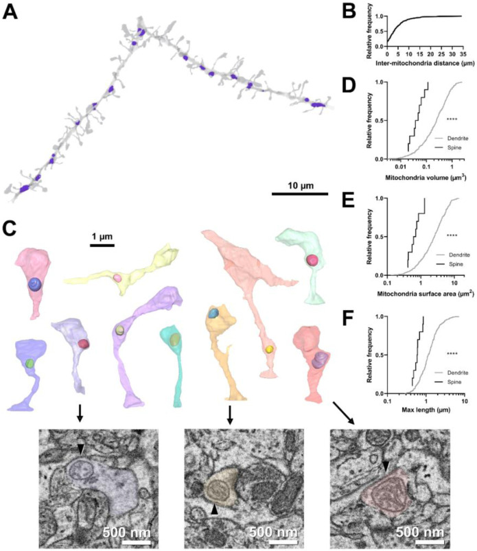
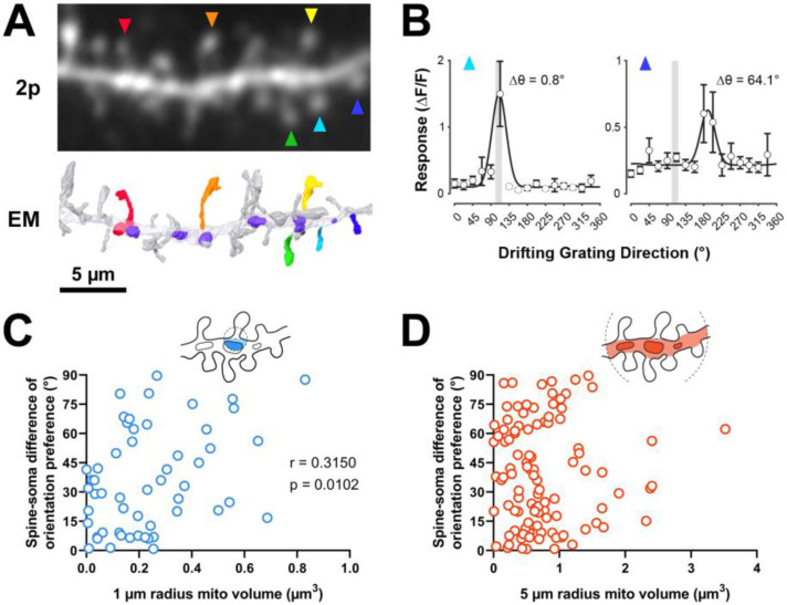
Postsynaptic mitochondria are critical to the development, plasticity, and maintenance of synaptic inputs. However, their relationship to synaptic structure and functional activity is unknown. We examined a correlative dataset from ferret visual cortex with two-photon calcium imaging of dendritic spines during visual stimulation and electron microscopy (EM) reconstructions of spine ultrastructure, investigating mitochondrial abundance near functionally- and structurally-characterized spines. Surprisingly, we found no correlation to structural measures of synaptic strength. Instead, we found that mitochondria are positioned near spines with orientation preferences that are dissimilar to the somatic preference. Additionally, we found that mitochondria are positioned near groups of spines with heterogeneous orientation preferences. For a subset of spines with mitochondrion in the head or neck, synapses were larger and exhibited greater selectivity to visual stimuli than those without a mitochondrion. Our data suggest mitochondria are not necessarily positioned to support the energy needs of strong spines, but rather support the structurally and functionally diverse inputs innervating the basal dendrites of cortical neurons.

摘要

突触后线粒体对于突触输入的发育、可塑性和维持至关重要。然而,它们与突触结构和功能活动之间的关系尚不清楚。我们研究了雪貂视觉皮层的一个相关数据集,该数据集包括视觉刺激期间树突棘的双光子钙成像以及棘突超微结构的电子显微镜(EM)重建,研究功能和结构特征明确的棘突附近的线粒体丰度。令人惊讶的是,我们发现与突触强度的结构测量值没有相关性。相反,我们发现线粒体位于棘突附近,其取向偏好与体细胞偏好不同。此外,我们发现线粒体位于取向偏好异质的棘突群附近。对于头部或颈部有线粒体的一部分棘突,突触比没有线粒体的突触更大,并且对视觉刺激表现出更高的选择性。我们的数据表明,线粒体不一定是为了支持强棘突的能量需求而定位的,而是支持支配皮层神经元基底树突的结构和功能多样的输入。

https://cdn.ncbi.nlm.nih.gov/pmc/blobs/fabb/10540538/38d6e6a55150/nihpp-2023.07.14.549063v2-f0006.jpg
https://cdn.ncbi.nlm.nih.gov/pmc/blobs/fabb/10540538/4c2fb88eb94c/nihpp-2023.07.14.549063v2-f0001.jpg
https://cdn.ncbi.nlm.nih.gov/pmc/blobs/fabb/10540538/4ab2b9dcbaaf/nihpp-2023.07.14.549063v2-f0002.jpg
https://cdn.ncbi.nlm.nih.gov/pmc/blobs/fabb/10540538/f6d12b3f12bb/nihpp-2023.07.14.549063v2-f0003.jpg
https://cdn.ncbi.nlm.nih.gov/pmc/blobs/fabb/10540538/6d127453c027/nihpp-2023.07.14.549063v2-f0004.jpg
https://cdn.ncbi.nlm.nih.gov/pmc/blobs/fabb/10540538/bedc942302f7/nihpp-2023.07.14.549063v2-f0005.jpg
https://cdn.ncbi.nlm.nih.gov/pmc/blobs/fabb/10540538/38d6e6a55150/nihpp-2023.07.14.549063v2-f0006.jpg

相似文献

1
Postsynaptic mitochondria are positioned to support functional diversity of dendritic spines.突触后线粒体的定位有助于支持树突棘的功能多样性。
bioRxiv. 2023 Sep 25:2023.07.14.549063. doi: 10.1101/2023.07.14.549063.
2
Postsynaptic mitochondria are positioned to support functional diversity of dendritic spines.突触后线粒体的定位支持树突棘功能的多样性。
Elife. 2023 Dec 7;12:RP89682. doi: 10.7554/eLife.89682.
3
Selective activation of BK channels in small-headed dendritic spines suppresses excitatory postsynaptic potentials.在小头树突棘中选择性激活大电导钙激活钾通道可抑制兴奋性突触后电位。
J Physiol. 2022 May;600(9):2165-2187. doi: 10.1113/JP282303. Epub 2022 Mar 9.
4
[Long-term high-fat diet's impact on synaptic plasticity in the visual cortex and hippocampus neurons: an experimental study].[长期高脂饮食对视觉皮层和海马神经元突触可塑性的影响:一项实验研究]
Zhonghua Yan Ke Za Zhi. 2023 Sep 11;59(9):730-739. doi: 10.3760/cma.j.cn112142-20221213-00633.
5
Calcium modeling of spine apparatus-containing human dendritic spines demonstrates an "all-or-nothing" communication switch between the spine head and dendrite.人树突棘内脊柱小体的钙成像表明,棘头和树突之间存在一种“全有或全无”的通讯开关。
PLoS Comput Biol. 2022 Apr 25;18(4):e1010069. doi: 10.1371/journal.pcbi.1010069. eCollection 2022 Apr.
6
Ultrastructure of dendritic spines: correlation between synaptic and spine morphologies.树突棘的超微结构:突触与棘形态之间的相关性。
Front Neurosci. 2007 Oct 15;1(1):131-43. doi: 10.3389/neuro.01.1.1.010.2007. eCollection 2007 Nov.
7
Orientation selectivity and the functional clustering of synaptic inputs in primary visual cortex.初级视觉皮层中突触输入的方向选择性与功能聚类
Nat Neurosci. 2016 Aug;19(8):1003-9. doi: 10.1038/nn.4323. Epub 2016 Jun 13.
8
Activity-dependent dendritic spine neck changes are correlated with synaptic strength.活性依赖的树突棘颈变化与突触强度相关。
Proc Natl Acad Sci U S A. 2014 Jul 15;111(28):E2895-904. doi: 10.1073/pnas.1321869111. Epub 2014 Jun 30.
9
Unique properties of dually innervated dendritic spines in pyramidal neurons of the somatosensory cortex uncovered by 3D correlative light and electron microscopy.通过三维共聚焦光镜和电子显微镜发现躯体感觉皮层锥体神经元中双重支配树突棘的独特性质。
PLoS Biol. 2021 Aug 24;19(8):e3001375. doi: 10.1371/journal.pbio.3001375. eCollection 2021 Aug.
10
Dendritic and somatic appendages of identified rubrospinal neurons of the cat.猫红核脊髓神经元的树突和胞体附属结构。
Neuroscience. 1987 Jul;22(1):113-30. doi: 10.1016/0306-4522(87)90202-8.