Abbate Jessica Maria, Palazzolo Simone, Ieni Antonio, Rapisarda Giuseppe Santi, Lanteri Giovanni
Department of Veterinary Sciences, University of Messina, Polo Universitario Annunziata, 98168 Messina, Italy.
University School for Advanced Studies IUSS Pavia, Piazza della Vittoria, 27100 Pavia, Italy.
Vet Sci. 2023 Jul 19;10(7):471. doi: 10.3390/vetsci10070471.
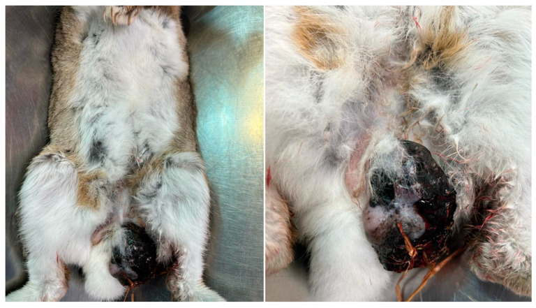
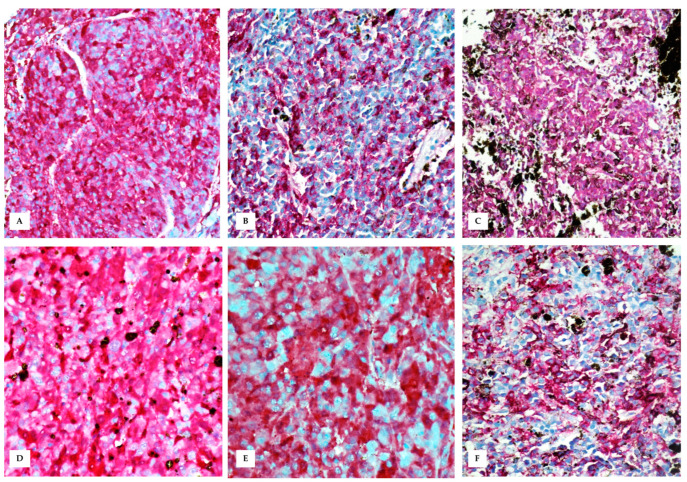
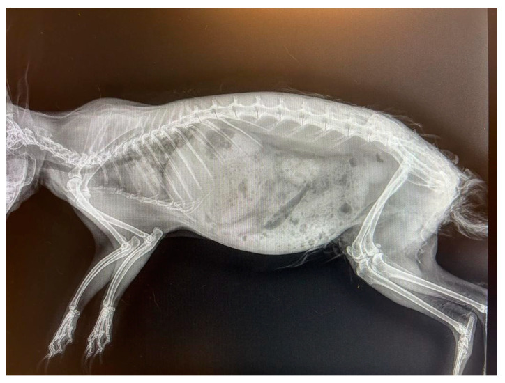
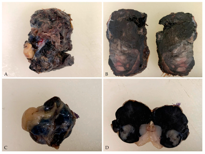
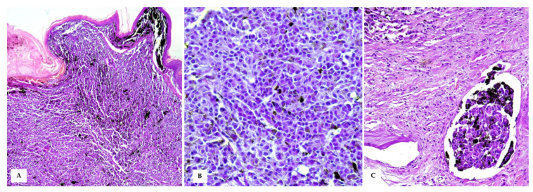
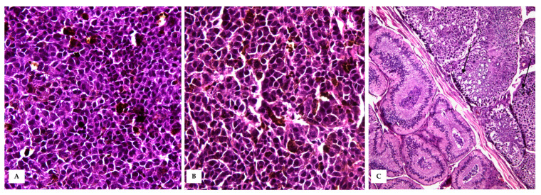
Melanocytic skin tumours have been rarely described in pet rabbits, and exposure to UV light in sparsely haired areas has been hypothesised to play a cancerogenic role. Here, we describe a case of cutaneous malignant melanoma arising from the skin of the scrotum in an 8-year-old male wild rabbit, with testicular metastases as an unusual metastatic site for melanoma reported in humans to date. The tumour was nearly 5 cm in size, firm, and highly pigmented, with multifocal superficial ulcerations and large areas of intratumoural necrosis. The adjacent testis was 1.5 cm, multinodular, and black, obscuring tissue morphology. Histologically, the dermis was expanded by an infiltrative, densely cellular neoplasm composed of nests and sheets of polygonal to spindle neoplastic melanocytes, supported by scant fibrovascular stroma. Neoplastic cells showed intermediate N/C ratio, moderate basophilic cytoplasm, often obscured by abundant brownish granular pigment, and eccentric nuclei with prominent nucleoli. Cellular pleomorphism and nuclear atypia were severe, and high mitotic activity was observed. Diffuse dermal lymphovascular invasion was also observed. The testis was delimited by a thin tunica albuginea, and the parenchyma was largely obscured in its morphology by densely packed neoplastic cells. Seminiferous tubules, lined with a thin basement membrane and containing neoplastic and scattered spermatogenic cells, were occasionally observed. Neoplastic cells within the skin and the testis were positive for HMB-45, Melan-A, and S-100. The growing popularity of rabbits as pets allows for a greater ability to accumulate data on the spontaneous occurrence of tumours in these animals. Furthermore, descriptions of the biological aspects of spontaneously occurring tumours may serve to improve current knowledge in animal species and humans in which the same neoplasm may occur.
黑素细胞性皮肤肿瘤在宠物兔中鲜有报道,有假说认为,在毛发稀疏区域暴露于紫外线下可能起到致癌作用。在此,我们描述了一例8岁雄性野兔阴囊皮肤发生的皮肤恶性黑色素瘤病例,睾丸转移是迄今为止人类黑色素瘤中罕见的转移部位。肿瘤大小近5厘米,质地坚硬,色素沉着明显,有多灶性浅表溃疡和大面积肿瘤内坏死。相邻睾丸大小为1.5厘米,呈多结节状,颜色发黑,组织形态难以辨认。组织学上,真皮被浸润性、细胞密集的肿瘤所占据,肿瘤由多边形至梭形的肿瘤性黑素细胞巢和片层组成,由少量纤维血管间质支持。肿瘤细胞显示中等核质比,胞质嗜碱性中等,常被大量棕褐色颗粒状色素遮盖,细胞核偏心,核仁突出。细胞多形性和核异型性严重,有较高的有丝分裂活性。还观察到弥漫性真皮淋巴管浸润。睾丸由一层薄的白膜界定,实质形态在很大程度上被密集堆积的肿瘤细胞遮盖。偶尔可见内衬薄基底膜、含有肿瘤细胞和散在生精细胞的生精小管。皮肤和睾丸内的肿瘤细胞HMB-45、Melan-A和S-100呈阳性。随着兔子作为宠物越来越受欢迎,我们有更多机会积累这些动物自发肿瘤发生情况的数据。此外,对自发发生肿瘤生物学方面的描述可能有助于增进我们对动物和人类中可能发生相同肿瘤的现有认识。