Department of Neurosurgery, Clinical Neuroscience Center, University Hospital Zurich, University of Zurich, Zurich, Switzerland.
Center for Microscopy and Image analysis, University of Zurich, Zurich, Switzerland.
Brain Pathol. 2023 Nov;33(6):e13189. doi: 10.1111/bpa.13189. Epub 2023 Jul 28.
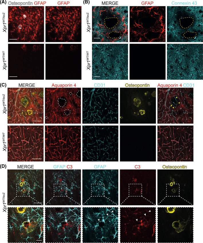
Calcification of the cerebral microvessels in the basal ganglia in the absence of systemic calcium and phosphate imbalance is a hallmark of primary familial brain calcification (PFBC), a rare neurodegenerative disorder. Mutation in genes encoding for sodium-dependent phosphate transporter 2 (SLC20A2), xenotropic and polytropic retrovirus receptor 1 (XPR1), platelet-derived growth factor B (PDGFB), platelet-derived growth factor receptor beta (PDGFRB), myogenesis regulating glycosidase (MYORG), and junctional adhesion molecule 2 (JAM2) are known to cause PFBC. Loss-of-function mutations in XPR1, the only known inorganic phosphate exporter in metazoans, causing dominantly inherited PFBC was first reported in 2015 but until now no studies in the brain have addressed whether loss of one functional allele leads to pathological alterations in mice, a commonly used organism to model human diseases. Here we show that mice heterozygous for Xpr1 (Xpr1 ) present with reduced inorganic phosphate levels in the cerebrospinal fluid and age- and sex-dependent growth of vascular calcifications in the thalamus. Vascular calcifications are surrounded by vascular basement membrane and are located at arterioles in the smooth muscle layer. Similar to previously characterized PFBC mouse models, vascular calcifications in Xpr1 mice contain bone matrix proteins and are surrounded by reactive astrocytes and microglia. However, microglial activation is not confined to calcified vessels but shows a widespread presence. In addition to vascular calcifications, we observed vessel tortuosity and transmission electron microscopy analysis revealed microangiopathy-endothelial swelling, phenotypic alterations in vascular smooth muscle cells, and thickening of the basement membrane.
基底节脑微血管的钙化,而不存在全身钙和磷酸盐失衡,是原发性家族性脑钙化(PFBC)的标志,这是一种罕见的神经退行性疾病。编码钠依赖性磷酸盐转运体 2(SLC20A2)、异嗜性和多瘤病毒受体 1(XPR1)、血小板衍生生长因子 B(PDGFB)、血小板衍生生长因子受体β(PDGFRB)、肌生成调节糖苷酶(MYORG)和连接黏附分子 2(JAM2)的基因突变被认为是导致 PFBC 的原因。XPR1 是后生动物中唯一已知的无机磷酸盐外排体,其功能丧失突变导致显性遗传性 PFBC 于 2015 年首次报道,但迄今为止,尚无研究在大脑中探讨是否失去一个功能性等位基因会导致小鼠的病理改变,而小鼠是一种常用于模拟人类疾病的常用生物体。在这里,我们发现 Xpr1(Xpr1)杂合子的小鼠在脑脊液中无机磷酸盐水平降低,并随着年龄和性别依赖性地在丘脑血管中出现钙化。血管钙化被血管基底膜包围,位于平滑肌层的小动脉中。与之前描述的 PFBC 小鼠模型相似,Xpr1 小鼠的血管钙化含有骨基质蛋白,并被反应性星形胶质细胞和小胶质细胞包围。然而,小胶质细胞的激活并不局限于钙化的血管,而是广泛存在。除了血管钙化,我们还观察到血管扭曲,透射电子显微镜分析显示微血管病变-内皮肿胀,血管平滑肌细胞表型改变,以及基底膜增厚。