Axe Neurosciences, Centre de Recherche du CHU de Québec-Université Laval, Québec, QC, Canada.
Department of Molecular Medicine, Université Laval, Québec City, QC, Canada.
J Neuroinflammation. 2022 Sep 27;19(1):235. doi: 10.1186/s12974-022-02595-8.
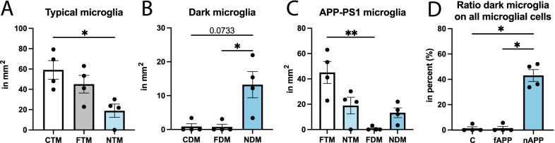
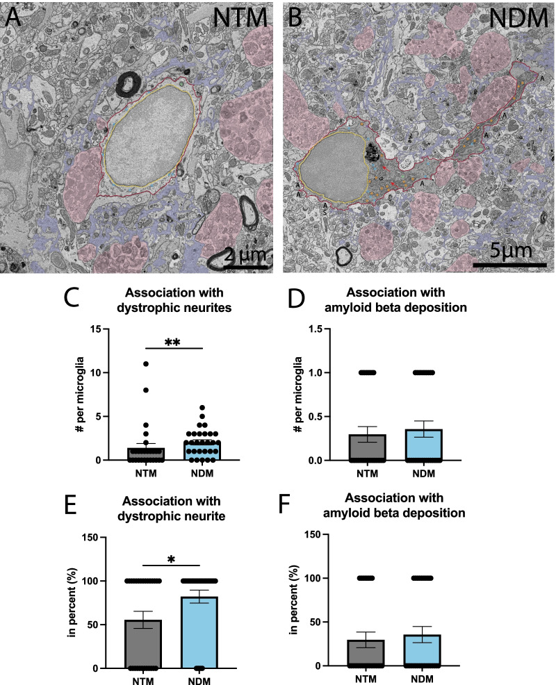
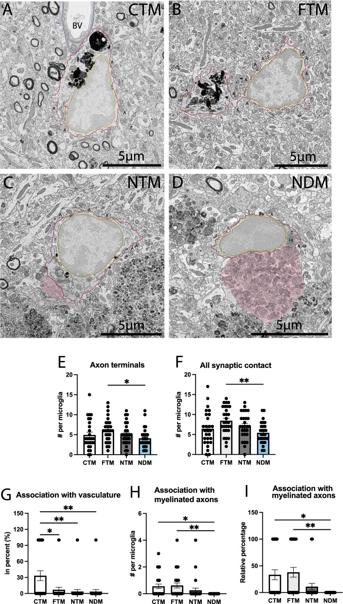
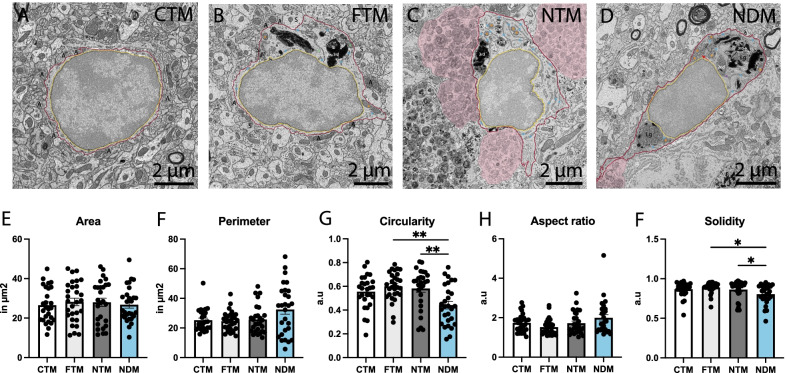
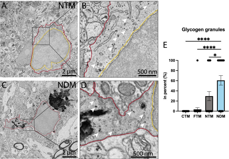
A diverse heterogeneity of microglial cells was previously described in Alzheimer's disease (AD) pathology, including dark microglia, a state characterized by ultrastructural markers of cellular stress. To provide novel insights into the roles of dark microglia during aging in the context of AD pathology, we performed a quantitative density and ultrastructural analysis of these cells using high-throughput scanning electron microscopy in the ventral hippocampus CA1 stratum lacunosum-moleculare of 20-month-old APP-PS1 vs C57BL/6J male mice. The density of dark microglia was significantly higher in APP-PS1 vs C57BL/6J mice, with these cells accounting for nearly half of all microglia observed near amyloid-beta (Aβ) plaques. This dark microglial state interacted more with dystrophic neurites compared to other APP-PS1 microglia and possessed glycogen granules, associated with a metabolic shift toward glycolysis, which provides the first ultrastructural evidence of their presence in microglia. Dark microglia were further observed in aging human post-mortem brain samples showing similar ultrastructural features as in mouse. Overall, our results provide a quantitative ultrastructural characterization of a microglial state associated with cellular stress (i.e., dark microglia) that is primarily restricted near Aβ plaques and dystrophic neurites. The presence of this microglial state in the aging human post-mortem brain is further revealed.
先前在阿尔茨海默病(AD)病理学中描述了多种异质性的小胶质细胞,包括暗小胶质细胞,其特征是存在细胞应激的超微结构标志物。为了深入了解暗小胶质细胞在 AD 病理学背景下衰老过程中的作用,我们使用高通量扫描电子显微镜对 20 月龄 APP-PS1 雄性小鼠和 C57BL/6J 雄性小鼠腹侧海马 CA1 腔隙分子层中的这些细胞进行了定量密度和超微结构分析。与 C57BL/6J 小鼠相比,APP-PS1 小鼠中暗小胶质细胞的密度明显更高,这些细胞几乎占所有靠近淀粉样β(Aβ)斑块的小胶质细胞的一半。与其他 APP-PS1 小胶质细胞相比,这种暗小胶质细胞状态与神经突营养不良的相互作用更多,并具有糖原颗粒,与糖酵解代谢转变有关,这为它们在小胶质细胞中的存在提供了第一个超微结构证据。在衰老的人类尸检脑组织样本中也观察到了暗小胶质细胞,其具有与小鼠相似的超微结构特征。总的来说,我们的研究结果提供了一种与细胞应激相关的小胶质细胞状态(即暗小胶质细胞)的定量超微结构特征描述,该状态主要局限于 Aβ 斑块和神经突营养不良附近。在衰老的人类尸检脑组织中存在这种小胶质细胞状态的证据进一步被揭示。