Department of Cardiology, Amsterdam UMC, Vrije Universiteit Amsterdam, Amsterdam Cardiovascular Sciences, Amsterdam, The Netherlands.
Department of Cardiothoracic Surgery, Amsterdam UMC, Amsterdam, The Netherlands.
Int J Cardiovasc Imaging. 2023 Sep;39(9):1753-1763. doi: 10.1007/s10554-023-02866-2. Epub 2023 May 31.
Left atrial (LA) sphericity is a novel, geometry-based parameter that has been used to visualize and quantify LA geometrical remodeling in patients with atrial fibrillation (AF). This study examined the association between LA sphericity, and LA longitudinal strain and strain rate measured by feature-tracking in AF patients.
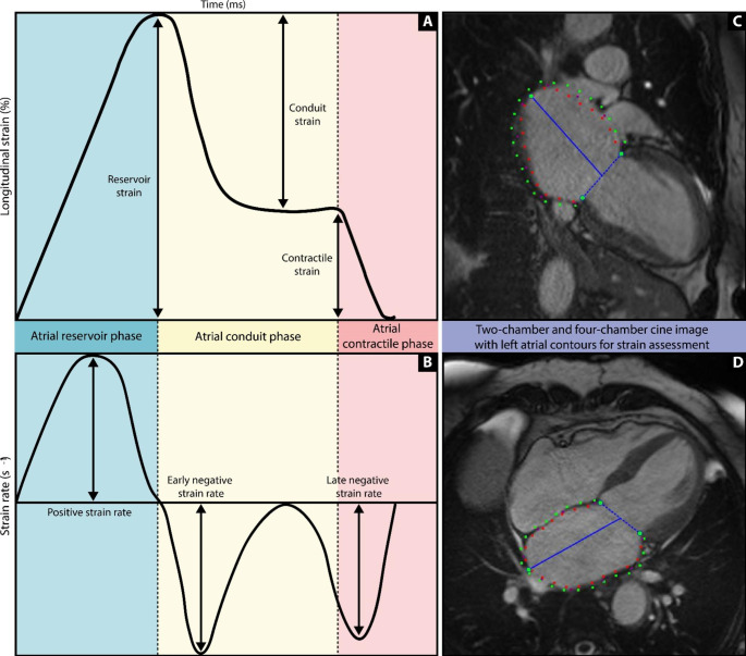
128 AF patients who underwent cardiovascular magnetic resonance (CMR) imaging in sinus rhythm prior to their pulmonary vein isolation (PVI) procedure were retrospectively analyzed. LA sphericity was calculated by segmenting the LA (excluding the pulmonary veins and the LA appendage) on a 3D contrast enhanced MR angiogram and comparing the resulting shape with a perfect sphere. LA global reservoir strain, conduit strain, contractile strain and corresponding strain rates were derived from cine images using feature-tracking. For statistical analysis, Pearson correlations, multivariable logistic regression analysis, and Student t-tests were used.
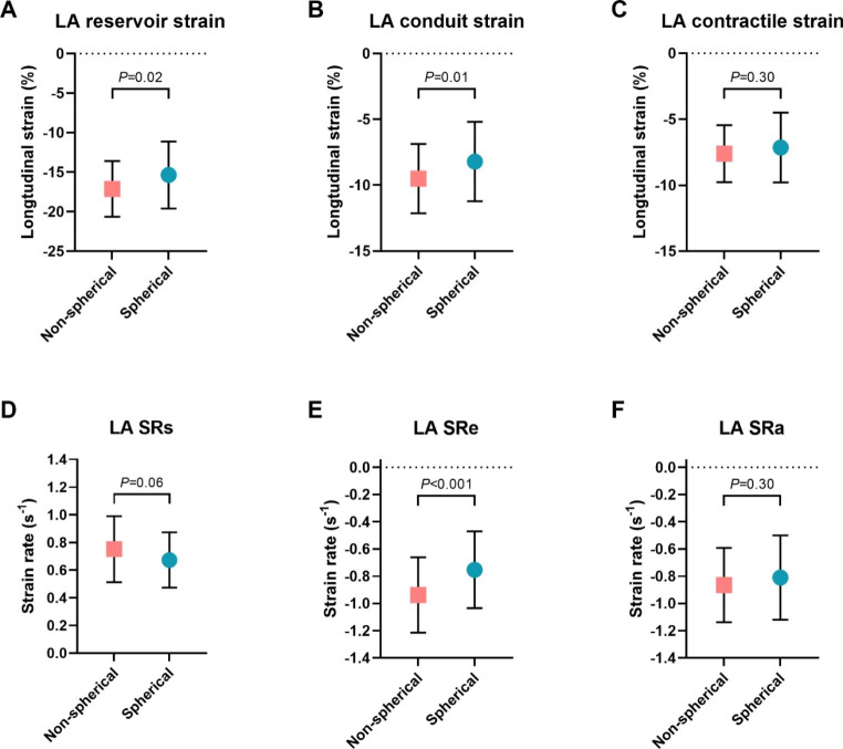
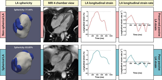
Patients with a spherical LA (dichotomized by the median value) had a lower reservoir strain and conduit strain compared to patients with a non-spherical LA (-15.4 ± 4.2% vs. -17.1 ± 3.5%, P = 0.02 and - 8.2 ± 3.0% vs. -9.5 ± 2.6%, P = 0.01, respectively). LA strain rate during early ventricular diastole was also different between both groups (-0.7 ± 0.3s vs. -0.9 ± 0.3s, P = 0.001). In contrast, no difference was found for LA contractile strain (-7.2 ± 2.6% vs. -7.6 ± 2.2%, P = 0.30).
LA passive strain is significantly impaired in AF patients with a spherical LA, though this relation was not independent from LA volume.
左心房(LA)的球形度是一种新的基于几何形状的参数,用于可视化和量化房颤(AF)患者的 LA 几何重塑。本研究探讨了 LA 球形度与 AF 患者通过特征追踪测量的 LA 纵向应变和应变率之间的关系。
回顾性分析了 128 例在肺静脉隔离(PVI)术前窦性心律下接受心血管磁共振(CMR)成像的 AF 患者。通过在 3D 对比增强磁共振血管造影上对 LA(不包括肺静脉和 LA 附件)进行分段,并将得到的形状与完美球体进行比较,计算 LA 球形度。使用特征追踪从电影图像中得出 LA 整体储层应变、导管应变、收缩应变和相应的应变率。统计分析采用 Pearson 相关、多变量逻辑回归分析和学生 t 检验。
球形 LA(按中位数二分)患者的储层应变和导管应变低于非球形 LA 患者(-15.4±4.2%比-17.1±3.5%,P=0.02 和-8.2±3.0%比-9.5±2.6%,P=0.01)。两组患者早期心室舒张期 LA 应变率也不同(-0.7±0.3s 比-0.9±0.3s,P=0.001)。相比之下,两组 LA 收缩应变无差异(-7.2±2.6%比-7.6±2.2%,P=0.30)。
LA 球形度的 AF 患者 LA 被动应变明显受损,但这种关系与 LA 容积无关。