• 文献检索
  • 文档翻译
  • 深度研究
  • 学术资讯
  • Suppr Zotero 插件Zotero 插件
  • 邀请有礼
  • 套餐&价格
  • 历史记录
应用&插件
Suppr Zotero 插件Zotero 插件浏览器插件Mac 客户端Windows 客户端微信小程序
定价
高级版会员购买积分包购买API积分包
服务
文献检索文档翻译深度研究API 文档MCP 服务
关于我们
关于 Suppr公司介绍联系我们用户协议隐私条款
关注我们

Suppr 超能文献

核心技术专利:CN118964589B侵权必究
粤ICP备2023148730 号-1Suppr @ 2026

文献检索

告别复杂PubMed语法,用中文像聊天一样搜索,搜遍4000万医学文献。AI智能推荐,让科研检索更轻松。

立即免费搜索

文件翻译

保留排版,准确专业,支持PDF/Word/PPT等文件格式,支持 12+语言互译。

免费翻译文档

深度研究

AI帮你快速写综述,25分钟生成高质量综述,智能提取关键信息,辅助科研写作。

立即免费体验

用于自动测量视神经鞘的商用即时超声评估。

Evaluation of commercially available point-of-care ultrasound for automated optic nerve sheath measurement.

作者信息

Moore Brad T, Osika Tom, Satterly Steven, Shah Shreyansh, Thirion Tim, Hampton Spencer, Aylward Stephen, Montgomery Sean

机构信息

Medical Computing, Kitware, Inc, Carrboro, NC, USA.

Surgical Critical Care, Duke University Health System, Durham, NC, USA.

出版信息

Ultrasound J. 2023 Aug 2;15(1):33. doi: 10.1186/s13089-023-00331-8.

DOI:10.1186/s13089-023-00331-8
PMID:37530991
原文链接:https://pmc.ncbi.nlm.nih.gov/articles/PMC10397168/
Abstract

BACKGROUND

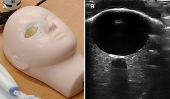
Measurement of the optic nerve sheath diameter (ONSD) via ultrasonography has been proposed as a non-invasive metric of intracranial pressure that may be employed during in-field patient triage. However, first responders are not typically trained to conduct sonographic exams and/or do not have access to an expensive ultrasound device. Therefore, for successful deployment of ONSD measurement in-field, we believe that first responders must have access to low-cost, portable ultrasound and be assisted by artificial intelligence (AI) systems that can automatically interpret the optic nerve sheath ultrasound scan. We examine the suitability of five commercially available, low-cost, portable ultrasound devices that can be combined with future artificial intelligence algorithms to reduce the training required for and cost of in-field optic nerve sheath diameter measurement. This paper is focused on the quality of the images generated by these low-cost probes. We report results of a clinician preference survey and compare with a lab analysis of three quantitative image quality metrics across devices. We also examine the suitability of the devices in a hypothetical far-forward deployment using operators unskilled in ultrasound, with the assumption of a future onboard AI video interpreter.

RESULTS

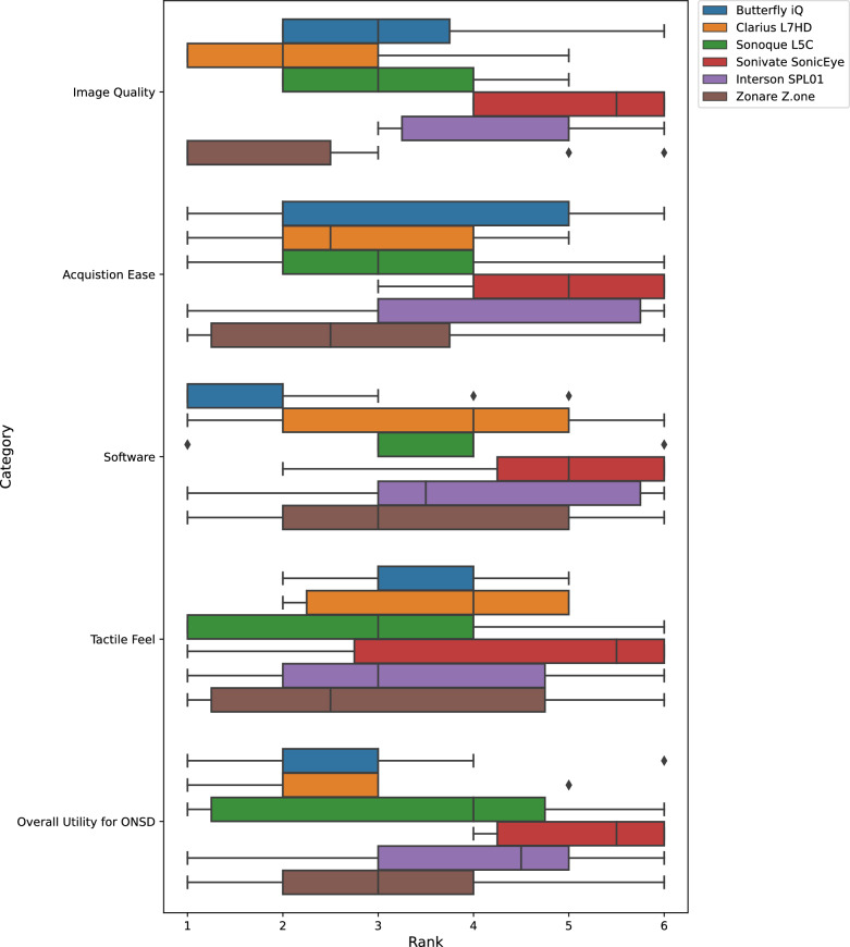
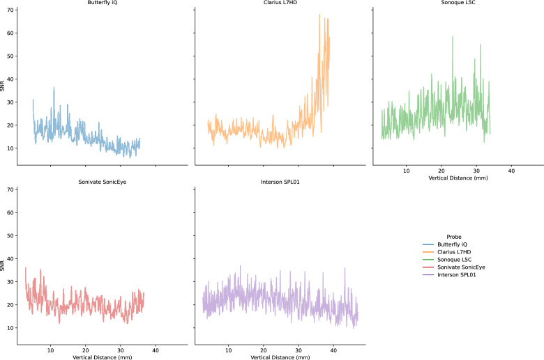
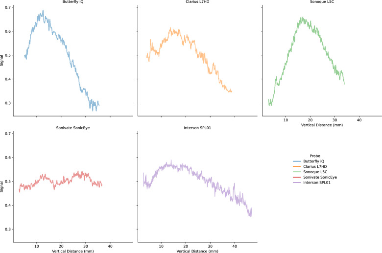
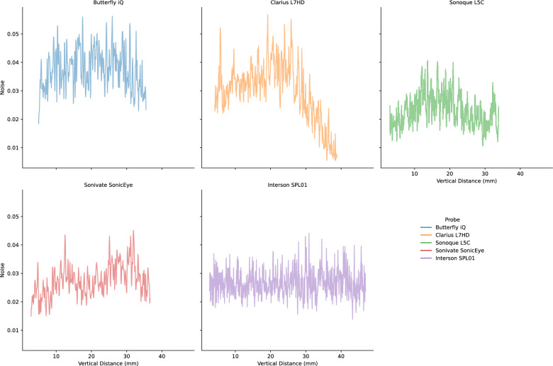
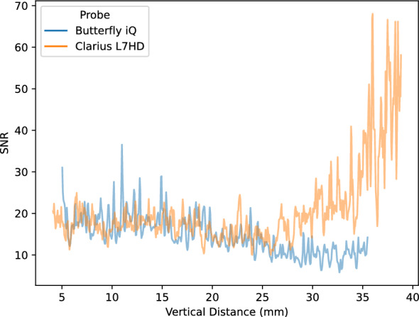
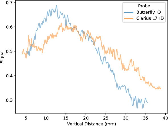
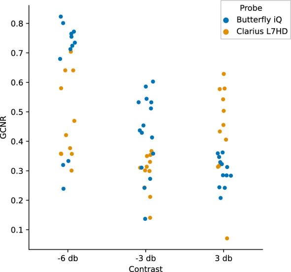
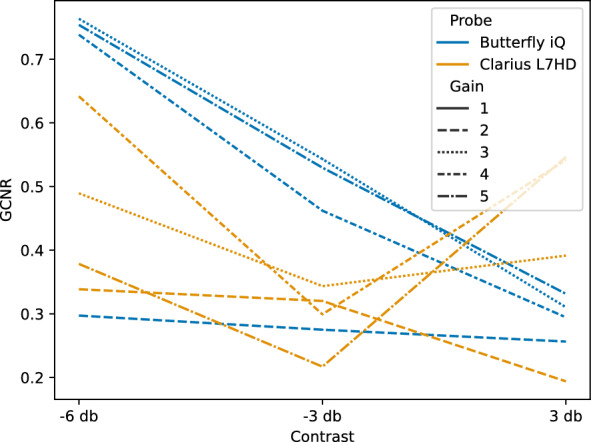
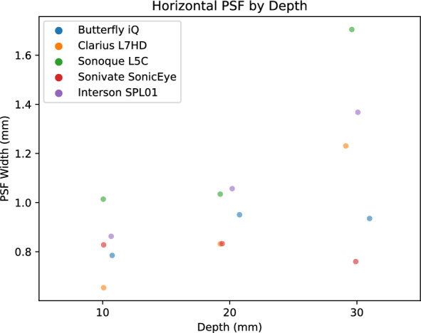
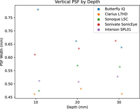
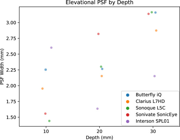
We find statistically significant differences in clinician ranking of the devices in the following categories: "Image Quality", "Ease of Acquisition", "Software", and "Overall ONSD". We show differences in signal-to-noise ratio, generalized contrast-to-noise ratio, point-spread function across the devices. These differences in image quality result in a statistically significant difference in manual ONSD measurement. Finally, we show that sufficiently wide transducers can capture the optic nerve sheath during blind (no visible B-mode) scans performed by operators unskilled in sonography.

CONCLUSIONS

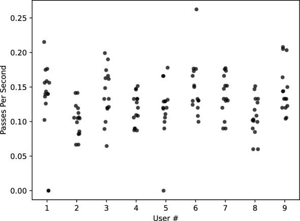
Ultrasound of the optic nerve sheath has the potential to be a convenient, non-invasive, point-of-injury or triage measure for elevated intracranial pressure in cases of traumatic brain injury. When transducer width is sufficient, briefly trained operators may obtain video sequences of the optic nerve sheath without guidance. This data suggest that unskilled operators are able to achieve the images needed for AI interpretation. However, we also show that image quality differences between ultrasound probes may influence manual ONSD measurements.

摘要

背景

通过超声测量视神经鞘直径(ONSD)已被提议作为一种颅内压的非侵入性指标,可用于现场患者分诊。然而,急救人员通常未接受过超声检查培训,和/或无法使用昂贵的超声设备。因此,为了在现场成功部署 ONSD 测量,我们认为急救人员必须能够使用低成本、便携式超声设备,并借助能够自动解读视神经鞘超声扫描结果的人工智能(AI)系统。我们研究了五种市售的低成本、便携式超声设备的适用性,这些设备可与未来的人工智能算法相结合,以降低现场测量视神经鞘直径所需的培训要求和成本。本文重点关注这些低成本探头生成的图像质量。我们报告了临床医生偏好调查的结果,并与对各设备的三个定量图像质量指标的实验室分析结果进行了比较。我们还在假设未来有车载人工智能视频解释器的情况下,研究了这些设备在假设的极前沿部署中对于不熟练超声操作的操作人员的适用性。

结果

我们发现临床医生在以下类别中对设备的排名存在统计学上的显著差异:“图像质量”、“采集难易程度”、“软件”和“总体 ONSD”。我们展示了各设备在信噪比、广义对比噪声比、点扩散函数方面的差异。这些图像质量差异导致手动测量 ONSD 时存在统计学上的显著差异。最后,我们表明,足够宽的换能器能够在不熟练超声操作的操作人员进行盲扫(无可见 B 模式)时捕捉到视神经鞘。

结论

视神经鞘超声有潜力成为创伤性脑损伤病例中颅内压升高的一种便捷、非侵入性的损伤点或分诊测量方法。当换能器宽度足够时,经过简短培训的操作人员无需指导即可获取视神经鞘的视频序列。这些数据表明,不熟练的操作人员能够获得人工智能解释所需图像。然而,我们也表明超声探头之间的图像质量差异可能会影响手动 ONSD 测量。

https://cdn.ncbi.nlm.nih.gov/pmc/blobs/2973/10397168/6f994b6adc59/13089_2023_331_Fig19_HTML.jpg
https://cdn.ncbi.nlm.nih.gov/pmc/blobs/2973/10397168/3b46874b0413/13089_2023_331_Fig1_HTML.jpg
https://cdn.ncbi.nlm.nih.gov/pmc/blobs/2973/10397168/1005651b15e6/13089_2023_331_Fig2_HTML.jpg
https://cdn.ncbi.nlm.nih.gov/pmc/blobs/2973/10397168/f5d4027762e6/13089_2023_331_Fig3_HTML.jpg
https://cdn.ncbi.nlm.nih.gov/pmc/blobs/2973/10397168/248e27464b27/13089_2023_331_Fig4_HTML.jpg
https://cdn.ncbi.nlm.nih.gov/pmc/blobs/2973/10397168/cdfd1d208854/13089_2023_331_Fig5_HTML.jpg
https://cdn.ncbi.nlm.nih.gov/pmc/blobs/2973/10397168/68207ee0b7af/13089_2023_331_Fig6_HTML.jpg
https://cdn.ncbi.nlm.nih.gov/pmc/blobs/2973/10397168/fd0a7d5a660e/13089_2023_331_Fig7_HTML.jpg
https://cdn.ncbi.nlm.nih.gov/pmc/blobs/2973/10397168/00f279c1f5ab/13089_2023_331_Fig8_HTML.jpg
https://cdn.ncbi.nlm.nih.gov/pmc/blobs/2973/10397168/21022e506413/13089_2023_331_Fig9_HTML.jpg
https://cdn.ncbi.nlm.nih.gov/pmc/blobs/2973/10397168/efd6ac2e26ef/13089_2023_331_Fig10_HTML.jpg
https://cdn.ncbi.nlm.nih.gov/pmc/blobs/2973/10397168/69768940b661/13089_2023_331_Fig11_HTML.jpg
https://cdn.ncbi.nlm.nih.gov/pmc/blobs/2973/10397168/263e7f2861a8/13089_2023_331_Fig12_HTML.jpg
https://cdn.ncbi.nlm.nih.gov/pmc/blobs/2973/10397168/485d223629ed/13089_2023_331_Fig13_HTML.jpg
https://cdn.ncbi.nlm.nih.gov/pmc/blobs/2973/10397168/093b7595c7f4/13089_2023_331_Fig14_HTML.jpg
https://cdn.ncbi.nlm.nih.gov/pmc/blobs/2973/10397168/c00579b48aba/13089_2023_331_Fig15_HTML.jpg
https://cdn.ncbi.nlm.nih.gov/pmc/blobs/2973/10397168/fe3cc2447ebc/13089_2023_331_Fig16_HTML.jpg
https://cdn.ncbi.nlm.nih.gov/pmc/blobs/2973/10397168/e86ae4e776b8/13089_2023_331_Fig17_HTML.jpg
https://cdn.ncbi.nlm.nih.gov/pmc/blobs/2973/10397168/ace1e89ecda3/13089_2023_331_Fig18_HTML.jpg
https://cdn.ncbi.nlm.nih.gov/pmc/blobs/2973/10397168/6f994b6adc59/13089_2023_331_Fig19_HTML.jpg

相似文献

1
Evaluation of commercially available point-of-care ultrasound for automated optic nerve sheath measurement.用于自动测量视神经鞘的商用即时超声评估。
Ultrasound J. 2023 Aug 2;15(1):33. doi: 10.1186/s13089-023-00331-8.
2
Noninvasive intracranial pressure assessment by optic nerve sheath diameter: Automated measurements as an alternative to clinician-performed measurements.通过视神经鞘直径进行无创颅内压评估:自动测量作为临床医生手动测量的替代方法。
Front Neurol. 2023 Feb 1;14:1064492. doi: 10.3389/fneur.2023.1064492. eCollection 2023.
3
Novel Algorithm for Automated Optic Nerve Sheath Diameter Measurement Using a Clustering Approach.一种使用聚类方法的自动视神经鞘直径测量的新算法。
Mil Med. 2021 Jan 25;186(Suppl 1):496-501. doi: 10.1093/milmed/usaa231.
4
Automatic Optic Nerve Measurement: A New Tool to Standardize Optic Nerve Assessment in Ultrasound B-Mode Images.自动视神经测量:一种标准化超声 B 模式图像视神经评估的新工具。
Ultrasound Med Biol. 2020 Jun;46(6):1533-1544. doi: 10.1016/j.ultrasmedbio.2020.01.034. Epub 2020 Mar 6.
5
Posterior Globe Depth for Optic Nerve Sheath Diameter Measurement in Ocular Ultrasound in Healthy Volunteers.健康志愿者眼超声视神经鞘直径测量的球后深度。
J Ultrasound Med. 2022 Oct;41(10):2425-2430. doi: 10.1002/jum.15925. Epub 2021 Dec 19.
6
Ratio of Optic Nerve Sheath Diameter to Eyeball Transverse Diameter by Ultrasound Can Predict Intracranial Hypertension in Traumatic Brain Injury Patients: A Prospective Study.超声测量视神经鞘直径与眼球横径的比值可预测颅脑外伤患者的颅内高压:一项前瞻性研究。
Neurocrit Care. 2020 Apr;32(2):478-485. doi: 10.1007/s12028-019-00762-z.
7
Development of a Deep Learning-Based System for Optic Nerve Characterization in Transorbital Ultrasound Images on a Multicenter Data Set.基于深度学习的眼眶超声图像视神经特征分析系统的多中心数据集研究
Ultrasound Med Biol. 2023 Sep;49(9):2060-2071. doi: 10.1016/j.ultrasmedbio.2023.05.011. Epub 2023 Jun 23.
8
Quality And Feasibility of Sonographic Measurement of the Optic Nerve Sheath Diameter to Estimate the Risk of Raised Intracranial Pressure After Traumatic Brain Injury in Prehospital Setting.院前环境下超声测量视神经鞘直径以评估创伤性脑损伤后颅内压升高风险的质量和可行性
Prehosp Emerg Care. 2019 Mar-Apr;23(2):277-283. doi: 10.1080/10903127.2018.1501444. Epub 2018 Aug 27.
9
Comparison of Three Point-of-Care Ultrasound Views and MRI Measurements for Optic Nerve Sheath Diameter: A Prospective Validity Study.三种床旁超声视图与磁共振成像测量视神经鞘直径的比较:一项前瞻性效度研究。
Neurocrit Care. 2020 Aug;33(1):173-181. doi: 10.1007/s12028-019-00881-7.
10
Optic nerve sheath diameter measurement: a means of detecting raised ICP in adult traumatic and non-traumatic neurosurgical patients.视神经鞘直径测量:一种检测成年创伤性和非创伤性神经外科患者颅内压升高的方法。
Am J Emerg Med. 2017 Jan;35(1):150-153. doi: 10.1016/j.ajem.2016.09.044. Epub 2016 Sep 23.

引用本文的文献

1
Comparing New Smartphone-Connected Handheld Ultrasound Device vs. Traditional Ultrasound in Vitreo-Retinal Disease Diagnosis.新型智能手机连接手持式超声设备与传统超声在玻璃体视网膜疾病诊断中的比较
Diagnostics (Basel). 2024 Sep 5;14(17):1961. doi: 10.3390/diagnostics14171961.
2
Inter-System Variability of Eight Different Handheld Ultrasound (HHUS) Devices-A Prospective Comparison of B-Scan Quality and Clinical Significance in Intensive Care.八种不同手持式超声(HHUS)设备的系统间变异性——重症监护中B超扫描质量及临床意义的前瞻性比较
Diagnostics (Basel). 2023 Dec 26;14(1):54. doi: 10.3390/diagnostics14010054.

本文引用的文献

1
The CLOSED protocol to assess optic nerve sheath diameter using color-Doppler: a comparison study in a cohort of idiopathic normal pressure hydrocephalus patients.使用彩色多普勒评估视神经鞘直径的CLOSED方案:特发性正常压力脑积水患者队列的比较研究
Ultrasound J. 2022 Oct 29;14(1):43. doi: 10.1186/s13089-022-00291-5.
2
Optic Nerve Sheath Diameter Ultrasound: A Non-Invasive Approach to Evaluate Increased Intracranial Pressure in Critically Ill Pediatric Patients.视神经鞘直径超声检查:一种评估危重症患儿颅内压升高的非侵入性方法。
Diagnostics (Basel). 2022 Mar 21;12(3):767. doi: 10.3390/diagnostics12030767.
3
Mean value of B-mode optic nerve sheath diameter as an indicator of increased intracranial pressure: a systematic review and meta-analysis.
作为颅内压升高指标的B超视神经鞘直径平均值:一项系统评价与Meta分析
Ultrasound J. 2021 Jul 2;13(1):35. doi: 10.1186/s13089-021-00235-5.
4
Novel Algorithm for Automated Optic Nerve Sheath Diameter Measurement Using a Clustering Approach.一种使用聚类方法的自动视神经鞘直径测量的新算法。
Mil Med. 2021 Jan 25;186(Suppl 1):496-501. doi: 10.1093/milmed/usaa231.
5
Automatic Optic Nerve Measurement: A New Tool to Standardize Optic Nerve Assessment in Ultrasound B-Mode Images.自动视神经测量:一种标准化超声 B 模式图像视神经评估的新工具。
Ultrasound Med Biol. 2020 Jun;46(6):1533-1544. doi: 10.1016/j.ultrasmedbio.2020.01.034. Epub 2020 Mar 6.
6
SciPy 1.0: fundamental algorithms for scientific computing in Python.SciPy 1.0:Python 中的科学计算基础算法。
Nat Methods. 2020 Mar;17(3):261-272. doi: 10.1038/s41592-019-0686-2. Epub 2020 Feb 3.
7
The Generalized Contrast-to-Noise Ratio: A Formal Definition for Lesion Detectability.广义对比噪声比:一种用于检测病变的正式定义。
IEEE Trans Ultrason Ferroelectr Freq Control. 2020 Apr;67(4):745-759. doi: 10.1109/TUFFC.2019.2956855. Epub 2019 Nov 29.
8
A Proposal for a New Protocol for Sonographic Assessment of the Optic Nerve Sheath Diameter: The CLOSED Protocol.《视神经鞘直径超声评估的新方案(CLOSED 方案)提案》
Neurocrit Care. 2020 Feb;32(1):327-332. doi: 10.1007/s12028-019-00853-x.
9
Optic nerve sheath diameter: present and future perspectives for neurologists and critical care physicians.视神经鞘直径:神经科医生和重症监护医师的现状和未来展望。
Neurol Sci. 2019 Dec;40(12):2447-2457. doi: 10.1007/s10072-019-04015-x. Epub 2019 Jul 31.
10
Diagnosis of elevated intracranial pressure in critically ill adults: systematic review and meta-analysis.诊断危重症成人颅内压升高:系统评价和荟萃分析。
BMJ. 2019 Jul 24;366:l4225. doi: 10.1136/bmj.l4225.