Division of Endocrinology, Diabetes and Metabolism, Department of Medicine, University of Miami Miller School of Medicine, Miami, FL, USA.
Section of Endocrinology and Metabolism, John W. Deming Department of Medicine, Tulane University School of Medicine, New Orleans, LA, USA; Southeast Louisiana Veterans Health Care System, New Orleans, LA, USA; Tulane Center of Excellence in Sex-Based Biology & Medicine, New Orleans, LA, USA.
Cell Rep. 2023 Aug 29;42(8):112913. doi: 10.1016/j.celrep.2023.112913. Epub 2023 Aug 1.
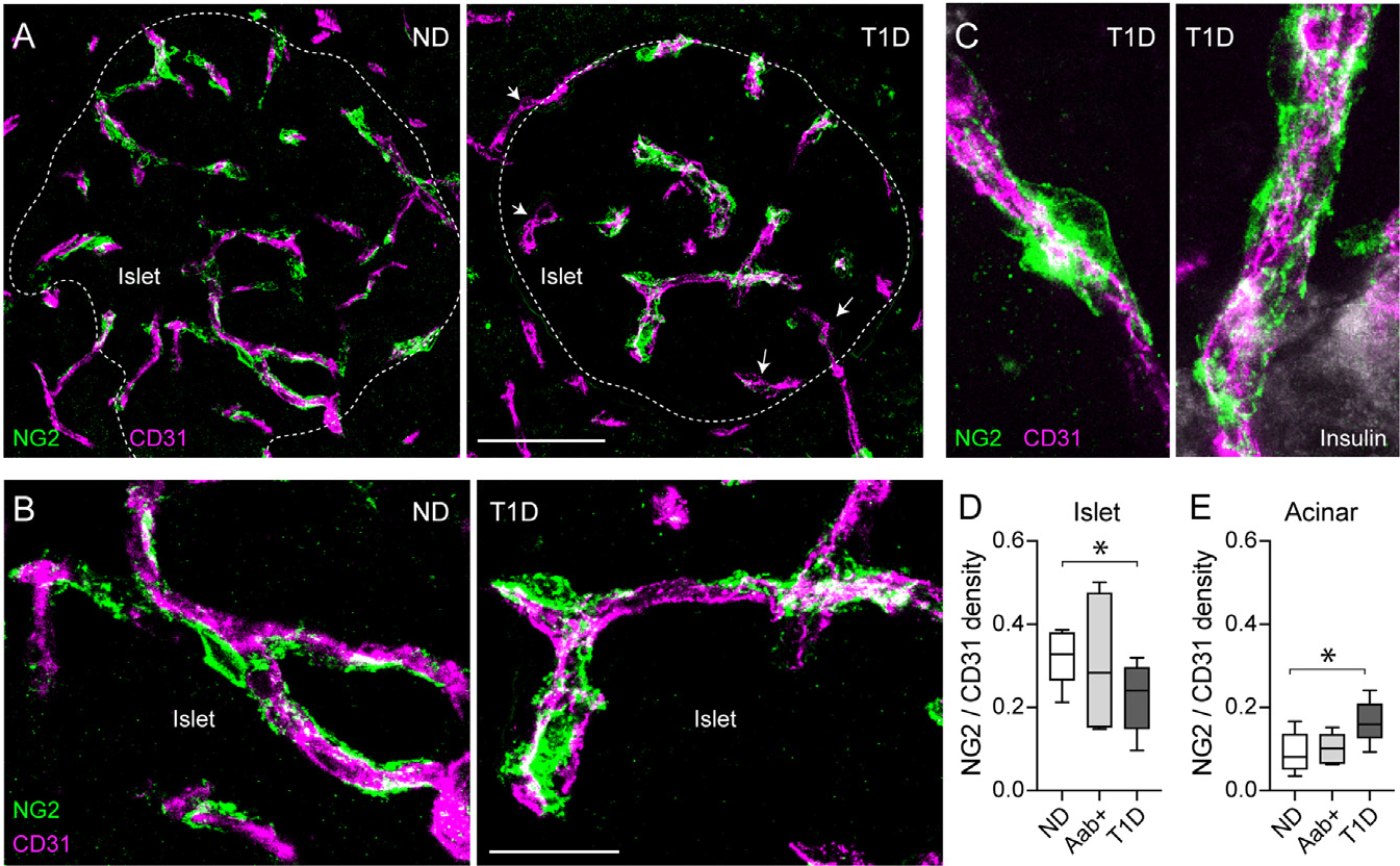
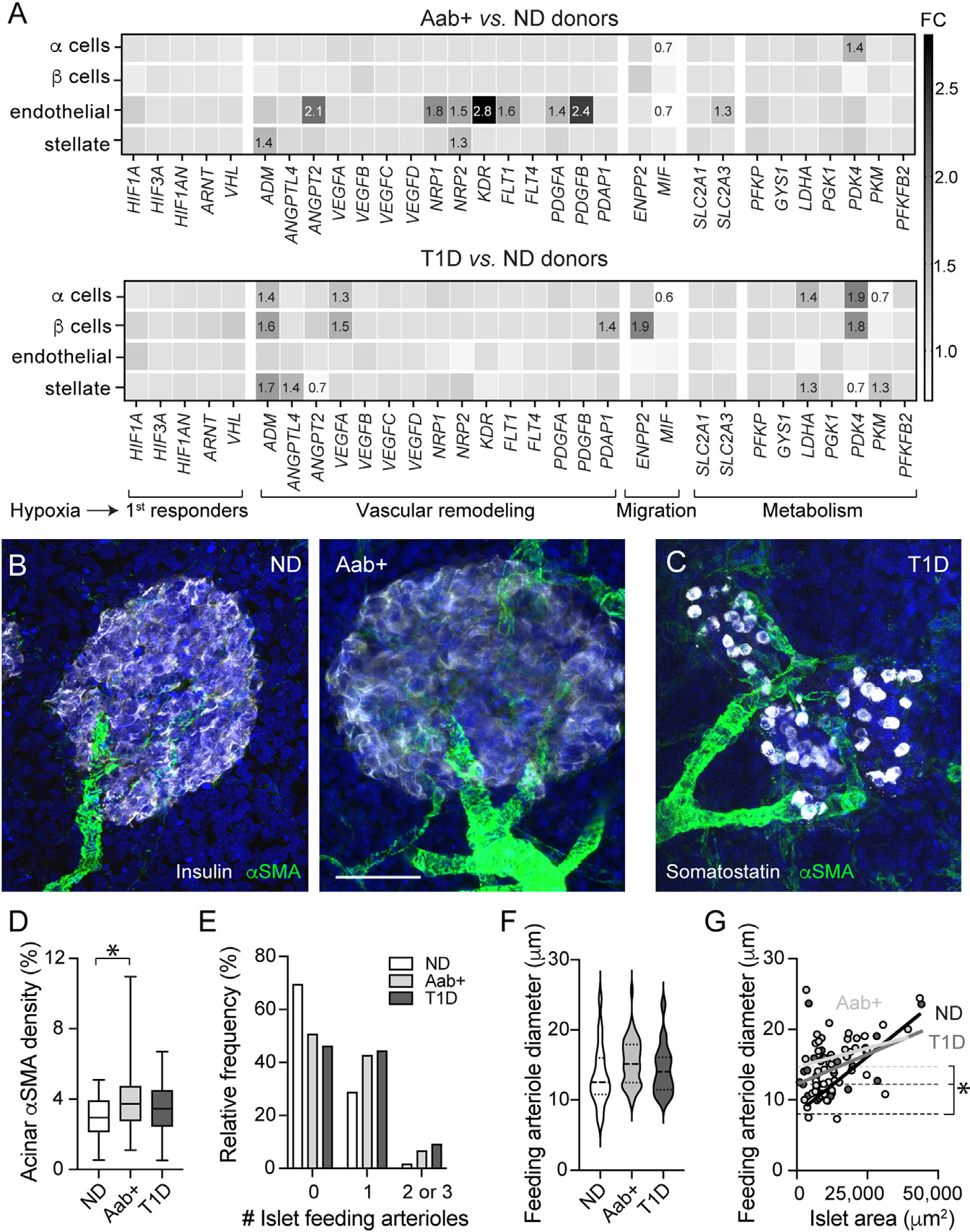
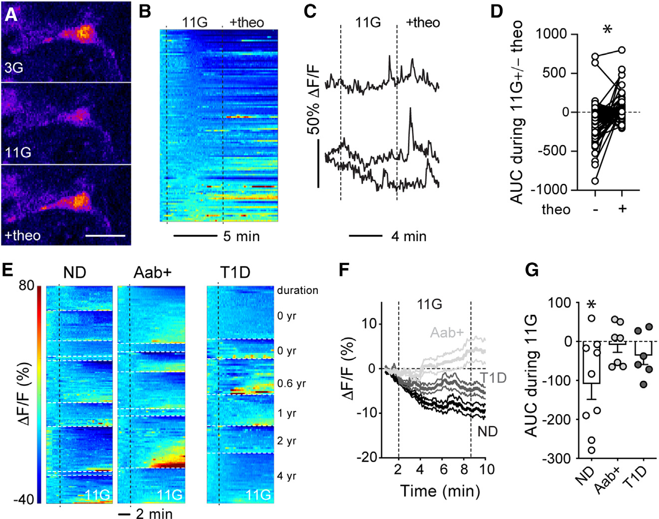
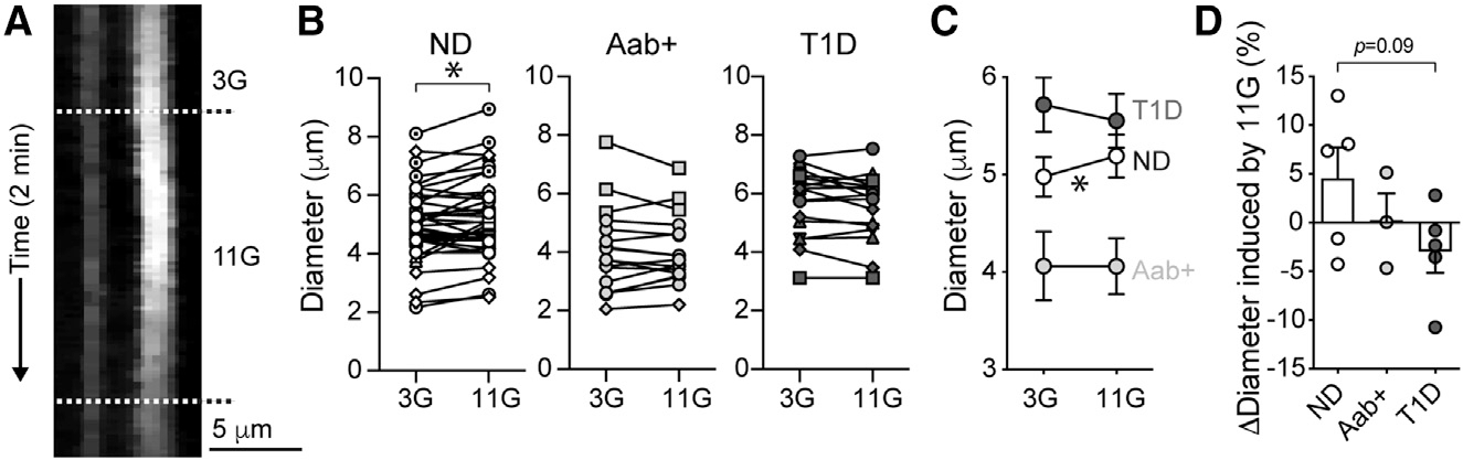
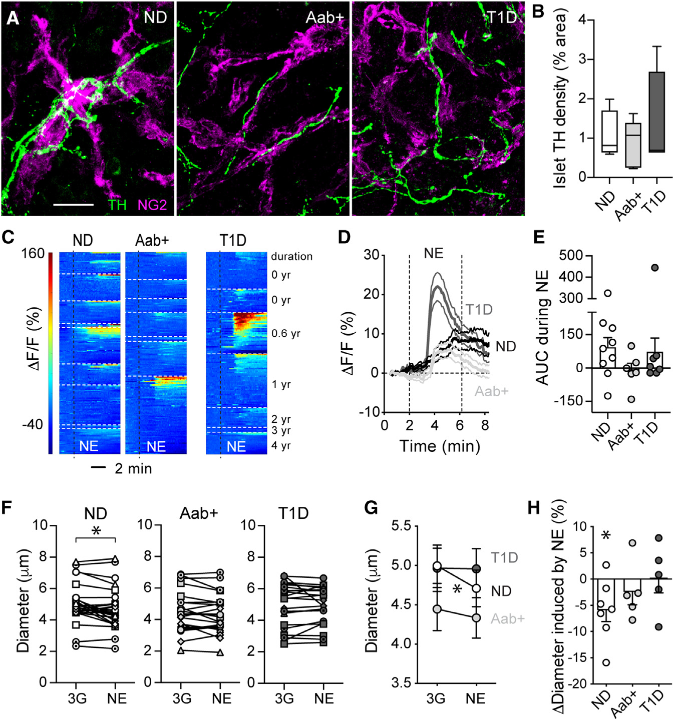
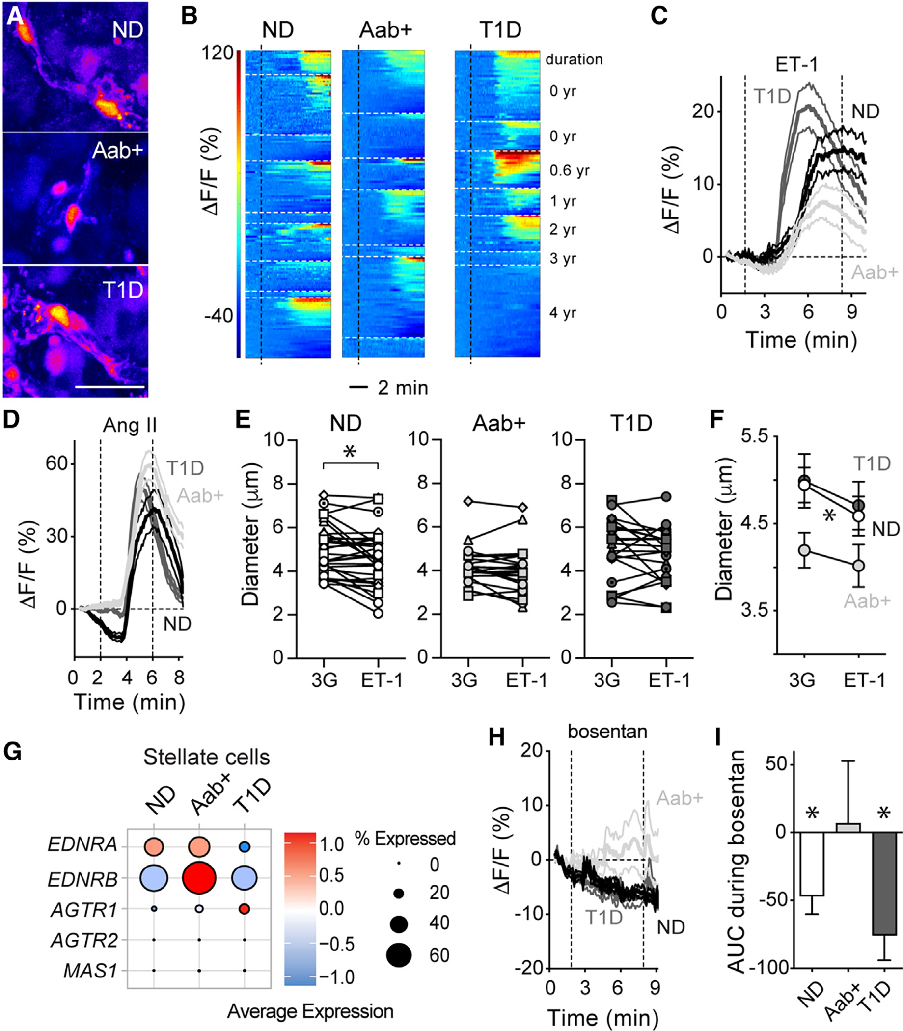
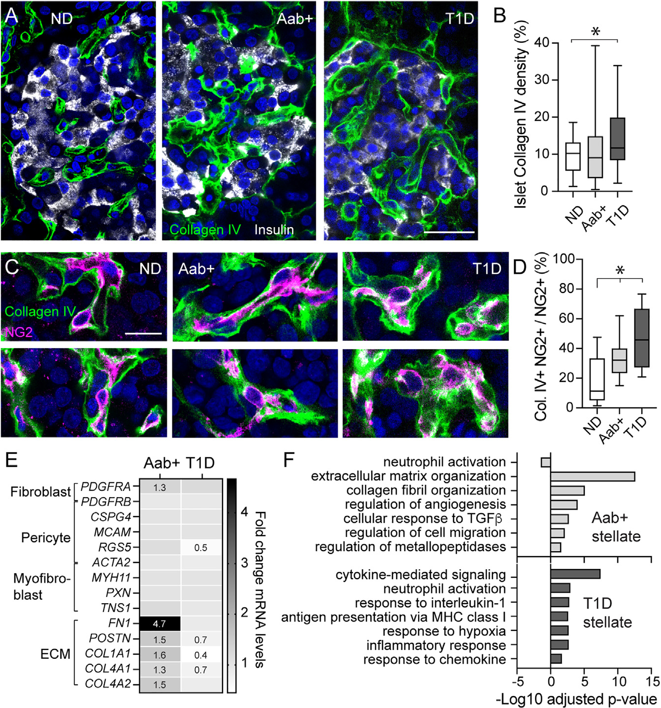
Pancreatic islets are endocrine organs that depend on their microvasculature to function. Along with endothelial cells, pericytes comprise the islet microvascular network. These mural cells are crucial for microvascular stability and function, but it is not known if/how they are affected during the development of type 1 diabetes (T1D). Here, we investigate islet pericyte density, phenotype, and function using living pancreas slices from donors without diabetes, donors with a single T1D-associated autoantibody (GADA+), and recent onset T1D cases. Our data show that islet pericyte and capillary responses to vasoactive stimuli are impaired early on in T1D. Microvascular dysfunction is associated with a switch in the phenotype of islet pericytes toward myofibroblasts. Using publicly available RNA sequencing (RNA-seq) data, we further found that transcriptional alterations related to endothelin-1 signaling and vascular and extracellular matrix (ECM) remodeling are hallmarks of single autoantibody (Aab)+ donor pancreata. Our data show that microvascular dysfunction is present at early stages of islet autoimmunity.
胰岛是依赖于其微血管系统发挥功能的内分泌器官。周细胞与内皮细胞一起构成胰岛微血管网络。这些壁细胞对于微血管的稳定性和功能至关重要,但尚不清楚它们在 1 型糖尿病 (T1D) 的发展过程中是如何受到影响的。在这里,我们使用来自无糖尿病供体、具有单个 T1D 相关自身抗体 (GADA+) 的供体和近期发生的 T1D 病例的活体胰腺切片,研究胰岛周细胞的密度、表型和功能。我们的数据表明,胰岛周细胞和毛细血管对血管活性刺激的反应在 T1D 早期就受到损害。微血管功能障碍与胰岛周细胞向肌成纤维细胞表型的转变有关。使用公开可用的 RNA 测序 (RNA-seq) 数据,我们进一步发现与内皮素-1 信号转导以及血管和细胞外基质 (ECM) 重塑相关的转录改变是单个自身抗体 (Aab)+供体胰腺的特征。我们的数据表明,微血管功能障碍存在于胰岛自身免疫的早期阶段。