Department of Veterinary Surgery, Cooperative Department of Veterinary Medicine, Faculty of Agriculture, Tokyo University of Agriculture and Technology, Fuchu 183-8509, Japan.
Laboratory of Veterinary Anatomy, Faculty of Veterinary Medicine, Okayama University of Science, Imabari 794-8555, Japan.
J Vet Sci. 2023 Jul;24(4):e50. doi: 10.4142/jvs.22290.
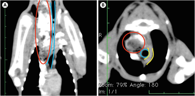
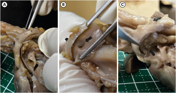
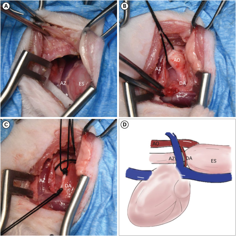
This paper reports the clinical findings and surgical treatment of feline right patent ductus arteriosus (RPDA) with a left aortic arch. A two-month-old female Maine Coon was referred for an investigation of regurgitation after weaning. RPDA with a left aortic arch was diagnosed based on the echocardiographic and computed tomography (CT) findings. A right-fourth intercostal thoracotomy was found to be an appropriate approach to the duct. Preoperative diagnosis is crucial and diagnostic imaging, including radiography, echocardiography, and cardiac CT examination, is essential for determining if the aortic arch is right or left.
本文报告了一例伴有左位主动脉弓的猫右动脉导管未闭(RPDA)的临床发现和手术治疗。一只两个月大的雌性缅因浣熊猫因断奶后出现反流而被转诊进行检查。根据超声心动图和计算机断层扫描(CT)结果诊断为 RPDA 伴左位主动脉弓。右第四肋间开胸术被认为是通向导管的合适途径。术前诊断至关重要,包括放射学、超声心动图和心脏 CT 检查在内的诊断影像学对于确定主动脉弓是右位还是左位是必要的。