Developmental Imaging, Murdoch Children's Research Institute, Royal Children's Hospital, Melbourne, 3052, Australia.
Neurodisability and Rehabilitation, Murdoch Children's Research Institute, Royal Children's Hospital, Melbourne, 3052, Australia.
Brain Struct Funct. 2023 Sep;228(7):1741-1754. doi: 10.1007/s00429-023-02682-3. Epub 2023 Aug 3.
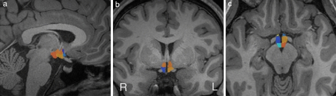
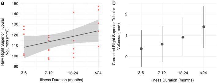
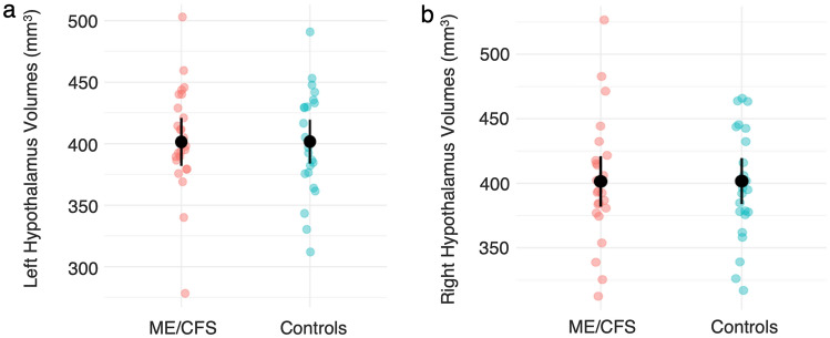
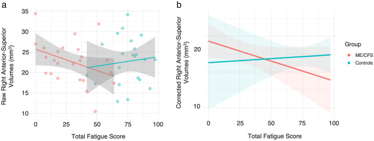
Adolescent Myalgic Encephalomyelitis/Chronic Fatigue Syndrome (ME/CFS) is a complex illness of unknown aetiology. Emerging theories suggest ME/CFS may reflect a progressive, aberrant state of homeostasis caused by disturbances within the hypothalamus, yet few studies have investigated this using magnetic resonance imaging in adolescents with ME/CFS. We conducted a volumetric analysis to investigate whether whole and regional hypothalamus volumes in adolescents with ME/CFS differed compared to healthy controls, and whether these volumes were associated with fatigue severity and illness duration. 48 adolescents (25 ME/CFS, 23 controls) were recruited. Lateralised whole and regional hypothalamus volumes, including the anterior-superior, superior tubular, posterior, anterior-inferior and inferior tubular subregions, were calculated from T1-weighted images. When controlling for age, sex and intracranial volume, Bayesian linear regression models revealed no evidence for differences in hypothalamus volumes between groups. However, in the ME/CFS group, a weak linear relationship between increased right anterior-superior volumes and fatigue severity was identified, which was absent in controls. In addition, Bayesian quantile regression revealed a likely-positive association between illness duration and right superior tubular volumes in the ME/CFS group. While these findings suggest overall comparability in regional and whole hypothalamus volumes between adolescents with ME/CFS and controls, preliminary evidence was identified to suggest greater fatigue severity and longer illness duration were associated with greater right anterior-superior and superior-tubular volumes, respectively. These regions contain the anterior and superior divisions of the paraventricular nucleus, involved in the neuroendocrine response to stress, suggesting involvement in ME/CFS pathophysiology. However, replication in a larger, longitudinal cohort is required.
青少年肌痛性脑脊髓炎/慢性疲劳综合征 (ME/CFS) 是一种病因不明的复杂疾病。新兴理论表明,ME/CFS 可能反映了由下丘脑内部紊乱引起的渐进性、异常的内稳态状态,但很少有研究使用磁共振成像来研究青少年 ME/CFS 中的这种情况。我们进行了一项容积分析,以调查青少年 ME/CFS 患者的整个和区域性下丘脑体积是否与健康对照组存在差异,以及这些体积是否与疲劳严重程度和疾病持续时间相关。招募了 48 名青少年(25 名 ME/CFS,23 名对照组)。从 T1 加权图像中计算了侧化的整个和区域性下丘脑体积,包括前上、上管状、后、前下和下管状亚区。在控制年龄、性别和颅内体积后,贝叶斯线性回归模型显示组间下丘脑体积无差异。然而,在 ME/CFS 组中,发现右侧前上体积增加与疲劳严重程度之间存在微弱的线性关系,而对照组则不存在。此外,贝叶斯分位数回归显示 ME/CFS 组中疾病持续时间与右侧上管状体积之间可能存在正相关。虽然这些发现表明青少年 ME/CFS 患者和对照组的区域性和整个下丘脑体积总体上具有可比性,但初步证据表明,更大的疲劳严重程度和更长的疾病持续时间与更大的右侧前上和上管状体积相关。这些区域包含参与应激神经内分泌反应的室旁核的前和上部分,表明其参与了 ME/CFS 的病理生理学。然而,需要在更大的纵向队列中进行复制。