Pais-Roldán Patricia, Yun Seong Dae, Shah N Jon
Institute of Neuroscience and Medicine 4, Medical Imaging Physics, Forschungszentrum Jülich, Jülich, Germany.
Institute of Neuroscience and Medicine 11, Molecular Neuroscience and Neuroimaging, Jülich Aachen Research Alliance, Forschungszentrum Jülich, Jülich, Germany.
Front Neuroimaging. 2022 May 4;1:869454. doi: 10.3389/fnimg.2022.869454. eCollection 2022.
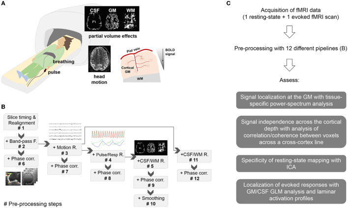
Over the past 30 years, brain function has primarily been evaluated non-invasively using functional magnetic resonance imaging (fMRI) with gradient-echo (GE) sequences to measure blood-oxygen-level-dependent (BOLD) signals. Despite the multiple advantages of GE sequences, e.g., higher signal-to-noise ratio, faster acquisitions, etc., their relatively inferior spatial localization compromises the routine use of GE-BOLD in laminar applications. Here, in an attempt to rescue the benefits of GE sequences, we evaluated the effect of existing pre-processing methods on the spatial localization of signals obtained with EPIK, a GE sequence that affords voxel volumes of 0.25 mm with near whole-brain coverage. The methods assessed here apply to both task and resting-state fMRI data assuming the availability of reconstructed magnitude and phase images.
在过去30年中,脑功能主要通过使用梯度回波(GE)序列的功能磁共振成像(fMRI)进行非侵入性评估,以测量血氧水平依赖(BOLD)信号。尽管GE序列具有多种优势,例如更高的信噪比、更快的采集速度等,但其相对较差的空间定位能力限制了GE-BOLD在层流应用中的常规使用。在此,为了挽救GE序列的优势,我们评估了现有预处理方法对使用EPIK获得的信号空间定位的影响,EPIK是一种GE序列,其体素体积为0.25 mm,几乎覆盖全脑。假设可获得重建的幅度和相位图像,这里评估的方法适用于任务态和静息态fMRI数据。