Department of Neurology, Washington University School of Medicine, St. Louis, Missouri, USA.
NeuroGenomics and Informatics Center, Washington University School of Medicine, St. Louis, Missouri, USA.
Alzheimers Dement. 2023 Oct;19(10):4488-4497. doi: 10.1002/alz.13377. Epub 2023 Aug 10.
Vascular damage in Alzheimer's disease (AD) has shown conflicting findings particularly when analyzing longitudinal data. We introduce white matter hyperintensity (WMH) longitudinal morphometric analysis (WLMA) that quantifies WMH expansion as the distance from lesion voxels to a region of interest boundary.
WMH segmentation maps were derived from 270 longitudinal fluid-attenuated inversion recovery (FLAIR) ADNI images. WLMA was performed on five data-driven WMH patterns with distinct spatial distributions. Amyloid accumulation was evaluated with WMH expansion across the five WMH patterns.
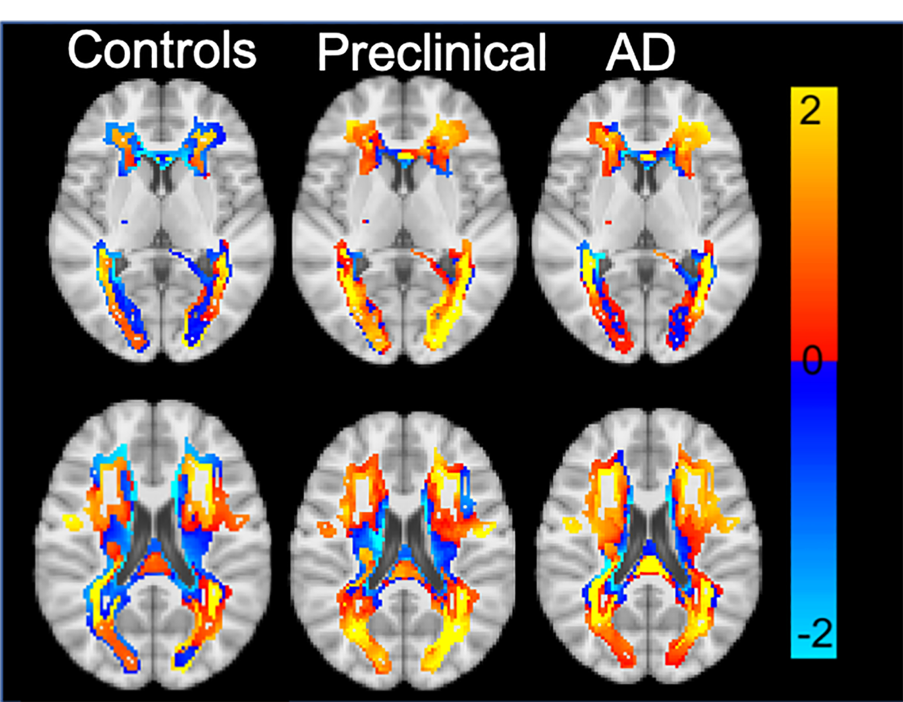
The preclinical group had significantly greater expansion in the posterior ventricular WM compared to controls. Amyloid significantly associated with frontal WMH expansion primarily within AD individuals. WLMA outperformed WMH volume changes for classifying AD from controls primarily in periventricular and posterior WMH.
These data support the concept that localized WMH expansion continues to proliferate with amyloid accumulation throughout the entirety of the disease in distinct spatial locations.
阿尔茨海默病(AD)中的血管损伤显示出相互矛盾的发现,特别是在分析纵向数据时。我们引入了脑白质高信号(WMH)的纵向形态计量分析(WLMA),该方法将 WMH 的扩展量化为病变体素与感兴趣区域边界之间的距离。
从 270 个纵向液体衰减反转恢复(FLAIR)ADNI 图像中得出了 WMH 分割图。WLMA 对具有不同空间分布的五个数据驱动的 WMH 模式进行了分析。用 WMH 扩展评估淀粉样蛋白的积累情况。
与对照组相比,临床前组在后脑室 WM 中的扩张明显更大。淀粉样蛋白与 AD 患者的额部 WMH 扩张显著相关。与 WMH 体积变化相比,WLMA 主要在脑室周围和后部 WMH 中对 AD 与对照组进行分类的效果更好。
这些数据支持了这样一种观点,即在疾病的整个过程中,淀粉样蛋白的积累会导致特定空间位置的局部性 WMH 扩张持续增生。