Department of Surgery, Collage of Medicine and Health Sciences, University of Gondar, Gondar, Ethiopia.
J Med Case Rep. 2023 Aug 15;17(1):348. doi: 10.1186/s13256-023-04078-7.
Calcified chronic subdural hematoma is a rare and infrequent diagnosis made in clinical practice according to the literature. Calcification of chronic subdural hematoma is found more frequently in children and young adults than in the aged. The proposed mechanism of calcification may involve poor circulation and absorption in the subdural space together with intravascular thrombosis and prolonged existence of the hematoma in the subdural space.
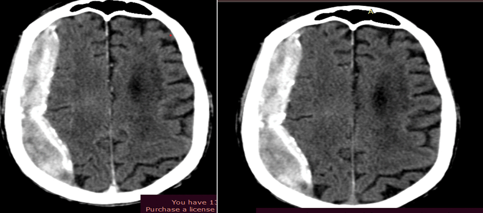
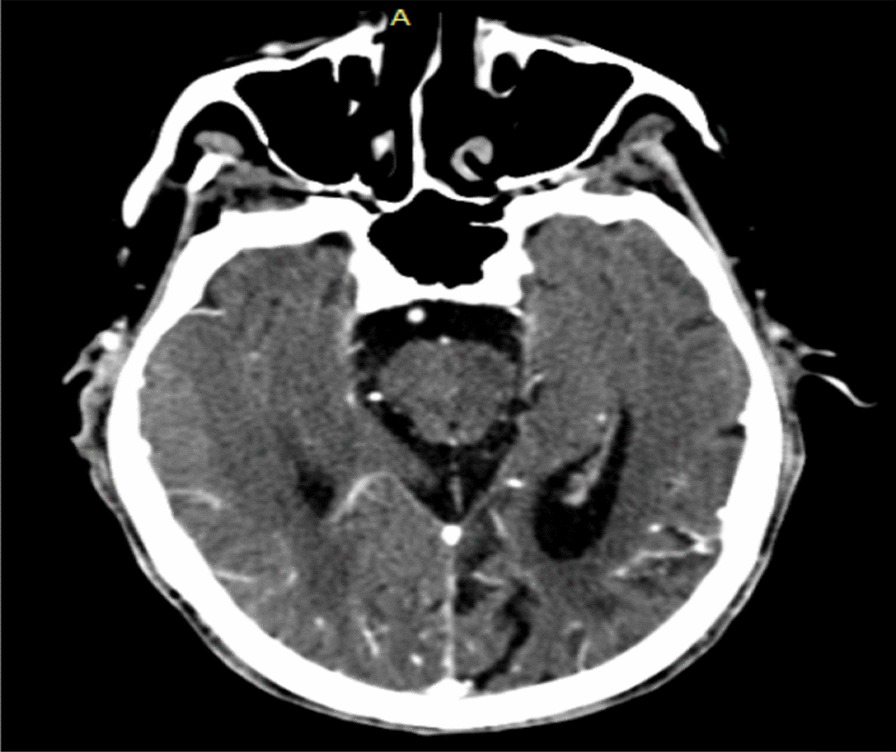
An 84-year-old Ethiopian male patient presented with progressive right-sided body weakness of 8-month duration. The weakness started in the right lower extremity and progressively involved the upper extremity. Associated with the above complaint, he had had also a globalized headache of the same duration. Pre- and post-contrast brain computed tomography scans showed a right hemispheric extra-axial collection that crossed the suture line, with a maximum depth of 2.3 cm. Subsequently, craniotomy and hematoma evacuation were carried out and the patient was discharged improved.
The most common symptom of calcified chronic subdural hematoma is headache followed by lethargy, confusion, memory impairment weakness, and seizures. A diminished level of consciousness is relatively common and motor deficits are usually manifested as hemiparesis or gait disturbance. Most calcified chronic subdural hematomas can be diagnosed by computed tomography or magnetic resonance imaging and differentiated from the usual chronic subdural hematoma by imaging studies and gross pathology. Surgical treatment is advised in symptomatic patients when feasible, and often results in neurological improvement. Here we presented a patient with an uncommon calcified chronic subdural hematoma, which was successfully evacuated, resulting in a good recovery.
根据文献记载,钙化性慢性硬脑膜下血肿在临床实践中是一种罕见且不常见的诊断。与老年人相比,儿童和青年更容易发生慢性硬脑膜下血肿钙化。钙化的可能机制包括硬脑膜下腔血液循环和吸收不良,以及血管内血栓形成和硬脑膜下血肿的长期存在。
一名 84 岁的埃塞俄比亚男性患者,右侧肢体无力进行性加重 8 个月。无力始于右下肢,并逐渐累及上肢。除上述症状外,他还伴有同样持续时间的全身性头痛。脑 CT 扫描(平扫+增强)显示右半球外轴状集合物,跨越缝线,最大深度为 2.3 厘米。随后进行了开颅血肿清除术,患者出院时病情好转。
钙化性慢性硬脑膜下血肿最常见的症状是头痛,其次是嗜睡、意识混乱、记忆力减退、乏力和癫痫。意识水平降低相对常见,运动缺陷通常表现为偏瘫或步态障碍。大多数钙化性慢性硬脑膜下血肿可通过计算机断层扫描或磁共振成像诊断,并通过影像学研究和大体病理学与常见的慢性硬脑膜下血肿相区别。当可行时,建议对有症状的患者进行手术治疗,通常可改善神经功能。在这里,我们介绍了一名患有罕见钙化性慢性硬脑膜下血肿的患者,该患者成功接受了清除术,恢复良好。