• 文献检索
  • 文档翻译
  • 深度研究
  • 学术资讯
  • Suppr Zotero 插件Zotero 插件
  • 邀请有礼
  • 套餐&价格
  • 历史记录
应用&插件
Suppr Zotero 插件Zotero 插件浏览器插件Mac 客户端Windows 客户端微信小程序
定价
高级版会员购买积分包购买API积分包
服务
文献检索文档翻译深度研究API 文档MCP 服务
关于我们
关于 Suppr公司介绍联系我们用户协议隐私条款
关注我们

Suppr 超能文献

核心技术专利:CN118964589B侵权必究
粤ICP备2023148730 号-1Suppr @ 2026

文献检索

告别复杂PubMed语法,用中文像聊天一样搜索,搜遍4000万医学文献。AI智能推荐,让科研检索更轻松。

立即免费搜索

文件翻译

保留排版,准确专业,支持PDF/Word/PPT等文件格式,支持 12+语言互译。

免费翻译文档

深度研究

AI帮你快速写综述,25分钟生成高质量综述,智能提取关键信息,辅助科研写作。

立即免费体验

与新冠后残留肺损伤最小的患者肺弥散功能变异性相关的定量CT指标

Quantitative CT Metrics Associated with Variability in the Diffusion Capacity of the Lung of Post-COVID-19 Patients with Minimal Residual Lung Lesions.

作者信息

Wen Han, Huapaya Julio A, Kanth Shreya M, Sun Junfeng, Matthew Brianna P, Lee Simone C, Do Michael, Chen Marcus Y, Malayeri Ashkan A, Suffredini Anthony F

机构信息

National Heart, Lung, and Blood Institute, National Institutes of Health, Bethesda, MD 20892, USA.

Critical Care Medicine Department, Clinical Center, National Institutes of Health, Bethesda, MD 20892, USA.

出版信息

J Imaging. 2023 Jul 26;9(8):150. doi: 10.3390/jimaging9080150.

DOI:10.3390/jimaging9080150
PMID:37623682
原文链接:https://pmc.ncbi.nlm.nih.gov/articles/PMC10455247/
Abstract

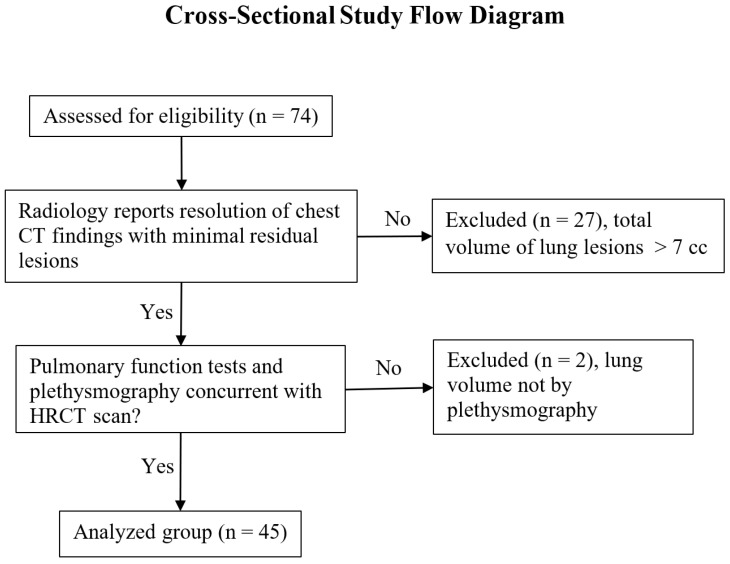
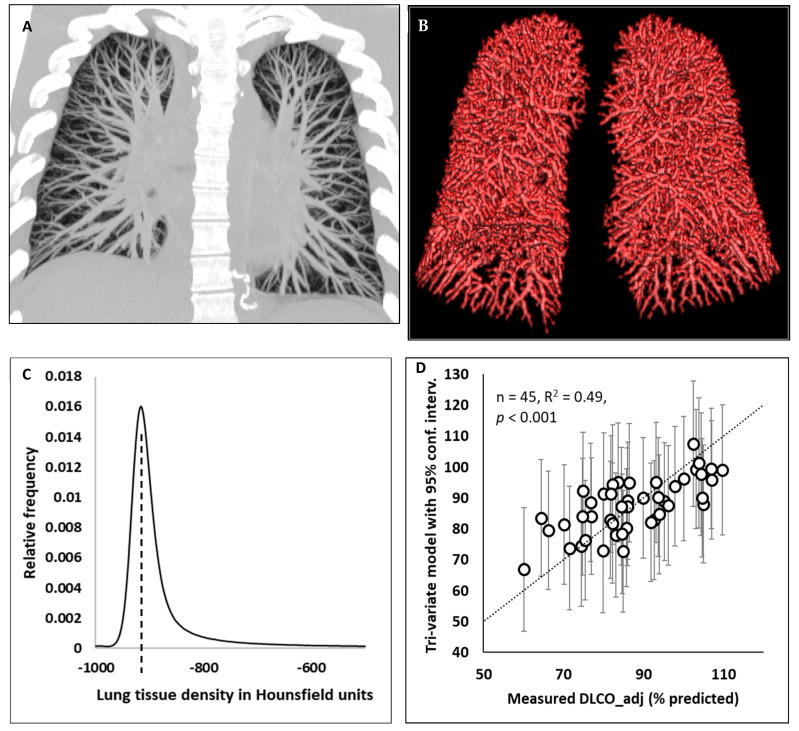
(1) Background: A reduction in the diffusion capacity of the lung for carbon monoxide is a prevalent longer-term consequence of COVID-19 infection. In patients who have zero or minimal residual radiological abnormalities in the lungs, it has been debated whether the cause was mainly due to a reduced alveolar volume or involved diffuse interstitial or vascular abnormalities. (2) Methods: We performed a cross-sectional study of 45 patients with either zero or minimal residual lesions in the lungs (total volume < 7 cc) at two months to one year post COVID-19 infection. There was considerable variability in the diffusion capacity of the lung for carbon monoxide, with 27% of the patients at less than 80% of the predicted reference. We investigated a set of independent variables that may affect the diffusion capacity of the lung, including demographic, pulmonary physiology and CT (computed tomography)-derived variables of vascular volume, parenchymal density and residual lesion volume. (3) Results: The leading three variables that contributed to the variability in the diffusion capacity of the lung for carbon monoxide were the alveolar volume, determined via pulmonary function tests, the blood vessel volume fraction, determined via CT, and the parenchymal radiodensity, also determined via CT. These factors explained 49% of the variance of the diffusion capacity, with values of 0.031, 0.005 and 0.018, respectively, after adjusting for confounders. A multiple-regression model combining these three variables fit the measured values of the diffusion capacity, with R = 0.70 and < 0.001. (4) Conclusions: The results are consistent with the notion that in some post-COVID-19 patients, after their pulmonary lesions resolve, diffuse changes in the vascular and parenchymal structures, in addition to a low alveolar volume, could be contributors to a lingering low diffusion capacity.

摘要

(1) 背景:肺一氧化碳弥散量降低是新冠病毒感染常见的长期后果。对于肺部影像学残留异常为零或极少的患者,其病因主要是肺泡容积减小还是涉及弥漫性间质或血管异常一直存在争议。(2) 方法:我们对45例新冠病毒感染后2个月至1年肺部残留病变为零或极少(总体积<7立方厘米)的患者进行了横断面研究。肺一氧化碳弥散量存在相当大的变异性,27%的患者低于预测参考值的80%。我们研究了一组可能影响肺弥散量的独立变量,包括人口统计学、肺生理学以及通过计算机断层扫描(CT)得出的血管容积、实质密度和残留病变体积等变量。(3) 结果:导致肺一氧化碳弥散量变异性的前三个变量分别是通过肺功能测试确定的肺泡容积、通过CT确定的血管容积分数以及同样通过CT确定的实质放射密度。在调整混杂因素后,这些因素分别解释了弥散量方差的49%,其系数分别为0.031、0.005和0.018。结合这三个变量的多元回归模型与弥散量测量值拟合良好,R = 0.70,P<0.001。(4) 结论:这些结果与以下观点一致,即在一些新冠病毒感染后的患者中,肺部病变消退后,除了肺泡容积较低外,血管和实质结构的弥漫性改变可能也是导致持续低弥散量的原因。

https://cdn.ncbi.nlm.nih.gov/pmc/blobs/fd49/10455247/653911e6b314/jimaging-09-00150-g002.jpg
https://cdn.ncbi.nlm.nih.gov/pmc/blobs/fd49/10455247/cf5c99d712c7/jimaging-09-00150-g001.jpg
https://cdn.ncbi.nlm.nih.gov/pmc/blobs/fd49/10455247/653911e6b314/jimaging-09-00150-g002.jpg
https://cdn.ncbi.nlm.nih.gov/pmc/blobs/fd49/10455247/cf5c99d712c7/jimaging-09-00150-g001.jpg
https://cdn.ncbi.nlm.nih.gov/pmc/blobs/fd49/10455247/653911e6b314/jimaging-09-00150-g002.jpg

相似文献

1
Quantitative CT Metrics Associated with Variability in the Diffusion Capacity of the Lung of Post-COVID-19 Patients with Minimal Residual Lung Lesions.与新冠后残留肺损伤最小的患者肺弥散功能变异性相关的定量CT指标
J Imaging. 2023 Jul 26;9(8):150. doi: 10.3390/jimaging9080150.
2
A cross-sectional study of quantitative CT measurements associated with the diffusion capacity of the lung in recovered COVID-19 patients with normalised chest CT.一项针对胸部CT已恢复正常的康复COVID-19患者中与肺弥散功能相关的定量CT测量的横断面研究。
medRxiv. 2022 Sep 15:2022.09.13.22279543. doi: 10.1101/2022.09.13.22279543.
3
Well-Aerated Lung and Mean Lung Density Quantified by CT at Discharge to Predict Pulmonary Diffusion Function 5 Months after COVID-19.通过CT量化出院时通气良好的肺和平均肺密度以预测COVID-19后5个月的肺扩散功能
Diagnostics (Basel). 2022 Nov 23;12(12):2921. doi: 10.3390/diagnostics12122921.
4
Lung Function, Radiological Findings and Biomarkers of Fibrogenesis in a Cohort of COVID-19 Patients Six Months After Hospital Discharge.一组新冠病毒肺炎患者出院六个月后的肺功能、影像学表现及纤维化生物标志物
Arch Bronconeumol. 2022 Feb;58(2):142-149. doi: 10.1016/j.arbres.2021.08.014. Epub 2021 Sep 3.
5
Diffusing capacity for carbon monoxide correlates best with tissue volume from quantitative CT scanning analysis.一氧化碳弥散量与定量 CT 扫描分析的组织体积相关性最好。
Chest. 2015 Jun;147(6):1485-1493. doi: 10.1378/chest.14-1693.
6
Long-lasting dyspnoea in patients otherwise clinically and radiologically recovered from COVID pneumonia: a probe for checking persisting disorders in capillary lung volume as a cause.新冠肺炎临床及影像学已康复患者的长期呼吸困难:探究肺毛细血管容量持续紊乱作为病因的可能性
Multidiscip Respir Med. 2022 Sep 30;17(1):875. doi: 10.4081/mrm.2022.875. eCollection 2022 Jan 12.
7
Results of quantitative chest-CT in chronic pulmonary graft-vs.-host disease (cGvHD) 3 years after allogeneic stem cell transplantation.异基因干细胞移植3年后慢性移植物抗宿主病(cGvHD)的胸部定量CT结果
J Thorac Dis. 2017 Aug;9(8):2521-2527. doi: 10.21037/jtd.2017.07.34.
8
Persistent Xe MRI Pulmonary and CT Vascular Abnormalities in Symptomatic Individuals with Post-acute COVID-19 Syndrome.持续性 Xe 肺部 MRI 和 CT 血管异常在急性 COVID-19 后综合征有症状个体中。
Radiology. 2022 Nov;305(2):466-476. doi: 10.1148/radiol.220492. Epub 2022 Jun 28.
9
Evaluation of computer-based computer tomography stratification against outcome models in connective tissue disease-related interstitial lung disease: a patient outcome study.基于计算机断层扫描分层对结缔组织病相关间质性肺病结局模型的评估:一项患者结局研究。
BMC Med. 2016 Nov 23;14(1):190. doi: 10.1186/s12916-016-0739-7.
10
A Follow-Up Study of Lung Function and Chest Computed Tomography at 6 Months after Discharge in Patients with Coronavirus Disease 2019.新冠肺炎患者出院后 6 个月时的肺功能和胸部计算机断层扫描随访研究。
Can Respir J. 2021 Feb 13;2021:6692409. doi: 10.1155/2021/6692409. eCollection 2021.

引用本文的文献

1
Prevalence and Cardiopulmonary Characteristics of Post-COVID Syndrome at a Hungarian Tertiary Referral Hospital.匈牙利一家三级转诊医院的新冠后综合征患病率及心肺特征
J Clin Med. 2025 Apr 10;14(8):2604. doi: 10.3390/jcm14082604.
2
Quantitative Computed Tomography Lung COVID Scores with Laboratory Markers: Utilization to Predict Rapid Progression and Monitor Longitudinal Changes in Patients with Coronavirus 2019 (COVID-19) Pneumonia.定量计算机断层扫描肺COVID评分与实验室指标:用于预测2019冠状病毒病(COVID-19)肺炎患者的快速进展并监测其纵向变化
Biomedicines. 2024 Jan 6;12(1):120. doi: 10.3390/biomedicines12010120.
3

本文引用的文献

1
European Respiratory Society statement on long COVID follow-up.欧洲呼吸学会关于新冠后症状长期随访的声明。
Eur Respir J. 2022 Aug 4;60(2). doi: 10.1183/13993003.02174-2021. Print 2022 Aug.
2
Association of abnormal pulmonary vasculature on CT scan for COVID-19 infection with decreased diffusion capacity in follow up: A retrospective cohort study.COVID-19 感染 CT 扫描中异常肺血管与随访中弥散能力下降的相关性:一项回顾性队列研究。
PLoS One. 2021 Oct 15;16(10):e0257892. doi: 10.1371/journal.pone.0257892. eCollection 2021.
3
Six-Month Pulmonary Impairment after Severe COVID-19: A Prospective, Multicentre Follow-Up Study.
Automated Segmentation and Measurements of Pulmonary Cysts in Lymphangioleiomyomatosis across Multiple CT Scanner Platforms over a Period of Two Decades.
二十年间跨多个CT扫描平台对淋巴管平滑肌瘤病中肺囊肿的自动分割与测量
Bioengineering (Basel). 2023 Oct 27;10(11):1255. doi: 10.3390/bioengineering10111255.
严重 COVID-19 后 6 个月的肺部损害:一项前瞻性、多中心随访研究。
Respiration. 2021;100(11):1078-1087. doi: 10.1159/000518141. Epub 2021 Aug 19.
4
Interstitial Lung Disease at High Resolution CT after SARS-CoV-2-Related Acute Respiratory Distress Syndrome According to Pulmonary Segmental Anatomy.根据肺段解剖结构分析2019冠状病毒病相关急性呼吸窘迫综合征后高分辨率CT表现的间质性肺疾病
J Clin Med. 2021 Sep 2;10(17):3985. doi: 10.3390/jcm10173985.
5
Residual ground glass opacities three months after Covid-19 pneumonia correlate to alteration of respiratory function: The post Covid M3 study.新冠肺炎后三个月的肺部磨玻璃影与呼吸功能改变相关:后新冠 M3 研究。
Respir Med. 2021 Aug;184:106435. doi: 10.1016/j.rmed.2021.106435. Epub 2021 May 15.
6
Medium-term impact of COVID-19 on pulmonary function, functional capacity and quality of life.COVID-19 对肺功能、功能能力和生活质量的中期影响。
Eur Respir J. 2021 Sep 16;58(3). doi: 10.1183/13993003.04015-2020. Print 2021 Sep.
7
Reduced Diffusion Capacity in COVID-19 Survivors.新冠康复者的弥散能力降低
Ann Am Thorac Soc. 2021 Jul;18(7):1253-1255. doi: 10.1513/AnnalsATS.202011-1452RL.
8
Cardiopulmonary recovery after COVID-19: an observational prospective multicentre trial.COVID-19 后心肺功能的恢复:一项观察性前瞻性多中心试验。
Eur Respir J. 2021 Apr 29;57(4). doi: 10.1183/13993003.03481-2020. Print 2021 Apr.
9
A prospective study of 12-week respiratory outcomes in COVID-19-related hospitalisations.一项针对 COVID-19 相关住院患者 12 周呼吸系统转归的前瞻性研究。
Thorax. 2021 Apr;76(4):402-404. doi: 10.1136/thoraxjnl-2020-216308. Epub 2020 Dec 3.
10
Caution in interpretation of abnormal carbon monoxide diffusion capacity in COVID-19 patients.注意解读 COVID-19 患者异常的一氧化碳扩散能力。
Eur Respir J. 2021 Jan 28;57(1). doi: 10.1183/13993003.03263-2020. Print 2021 Jan.