• 文献检索
  • 文档翻译
  • 深度研究
  • 学术资讯
  • Suppr Zotero 插件Zotero 插件
  • 邀请有礼
  • 套餐&价格
  • 历史记录
应用&插件
Suppr Zotero 插件Zotero 插件浏览器插件Mac 客户端Windows 客户端微信小程序
定价
高级版会员购买积分包购买API积分包
服务
文献检索文档翻译深度研究API 文档MCP 服务
关于我们
关于 Suppr公司介绍联系我们用户协议隐私条款
关注我们

Suppr 超能文献

核心技术专利:CN118964589B侵权必究
粤ICP备2023148730 号-1Suppr @ 2026

文献检索

告别复杂PubMed语法,用中文像聊天一样搜索,搜遍4000万医学文献。AI智能推荐,让科研检索更轻松。

立即免费搜索

文件翻译

保留排版,准确专业,支持PDF/Word/PPT等文件格式,支持 12+语言互译。

免费翻译文档

深度研究

AI帮你快速写综述,25分钟生成高质量综述,智能提取关键信息,辅助科研写作。

立即免费体验

在2型糖尿病小鼠模型中,神经视网膜的功能和结构变化伴随着线粒体功能障碍。

Functional and structural changes in the neuroretina are accompanied by mitochondrial dysfunction in a type 2 diabetic mouse model.

作者信息

Lam Christie Hang-I, Zou Bing, Chan Henry Ho-Lung, Tse Dennis Yan-Yin

机构信息

School of Optometry, The Hong Kong Polytechnic University, Hong Kong, SAR, China.

Centre for Eye and Vision Research Limited (CEVR), Hong Kong, SAR, China.

出版信息

Eye Vis (Lond). 2023 Sep 1;10(1):37. doi: 10.1186/s40662-023-00353-2.

DOI:10.1186/s40662-023-00353-2
PMID:37653465
原文链接:https://pmc.ncbi.nlm.nih.gov/articles/PMC10472703/
Abstract

BACKGROUND

Diabetic retinopathy (DR), one of the leading causes of blindness and vision impairment, is suggested to exhibit functional and structural changes in retinal neurons as the earliest manifestation, which could be used to predict the progression of related angiopathy. While neural function and survival rely on proper mitochondrial function, and a growing body of literature has supported the role of mitochondrial dysfunction in the development of DR, how diabetes affects mitochondrial function in retinal tissue remains elusive. This study primarily aimed to investigate mitochondrial functional changes in a diabetic rodent model. We also characterized the early DR phenotype, in particular, neurodegeneration.

METHODS

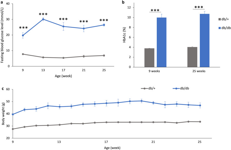
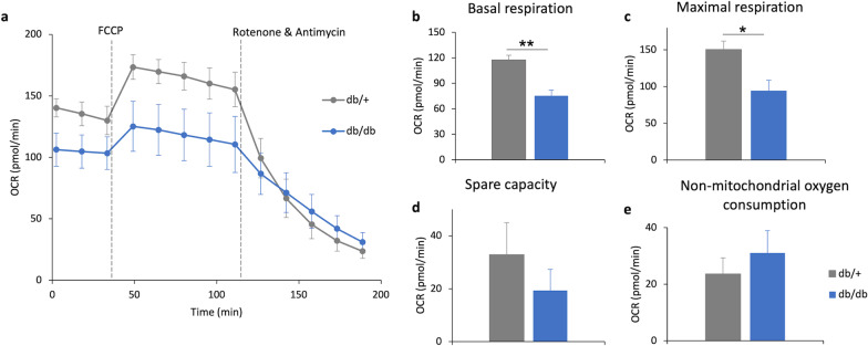
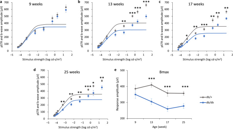
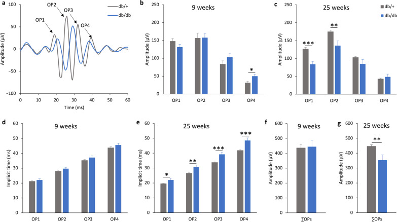
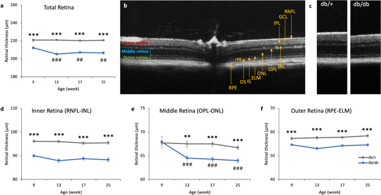
C57BLKsJ-db/db (db/db) mice (a type 2 diabetic mouse model) were used with their normoglycemic heterozygous littermates (db/+) serving as controls. Longitudinal changes in retinal function and morphology were assessed with electroretinography (ERG) and optical coherence tomography (OCT), respectively, at 9, 13, 17, and 25 weeks of age. At 25 weeks, the retinas were harvested for immunohistochemistry and ex vivo mitochondrial bioenergetics.

RESULTS

Decreased ERG responses were observed in db/db mice as early as 13 weeks of age. OCT revealed that db/db mice had significantly thinner retinas than the controls. Immunohistochemistry showed that the retinas of the db/db mice at 25 weeks were thinner at the outer and inner nuclear layers, with lower photoreceptor and cone cell densities compared with the db/+ mice. The number of rod-bipolar cell dendritic boutons and axon terminals was significantly reduced in db/db mice relative to the db/+ mice, suggesting that diabetes may lead to compromised synaptic connectivity. More importantly, the retinas of db/db mice had weaker mitochondrial functions than the controls.

CONCLUSIONS

Our longitudinal data suggest that diabetes-induced functional deterioration and morphological changes were accompanied by reduced mitochondrial function in the retina of db/db mice. These findings suggest that mitochondrial dysfunction may be a contributing factor triggering the development of DR. While the underlying mechanistic cause remains elusive, the db/db mice could be a useful animal model for testing potential treatment regimens targeting neurodegeneration in DR.

摘要

背景

糖尿病视网膜病变(DR)是导致失明和视力损害的主要原因之一,有研究表明,视网膜神经元的功能和结构变化是其最早的表现,可用于预测相关血管病变的进展。虽然神经功能和存活依赖于正常的线粒体功能,且越来越多的文献支持线粒体功能障碍在DR发生发展中的作用,但糖尿病如何影响视网膜组织中的线粒体功能仍不清楚。本研究主要旨在调查糖尿病啮齿动物模型中的线粒体功能变化。我们还对早期DR表型进行了特征描述,尤其是神经退行性变。

方法

使用C57BLKsJ-db/db(db/db)小鼠(一种2型糖尿病小鼠模型),其血糖正常的杂合子同窝小鼠(db/+)作为对照。分别在9、13、17和25周龄时,通过视网膜电图(ERG)和光学相干断层扫描(OCT)评估视网膜功能和形态的纵向变化。在25周时,摘取视网膜用于免疫组织化学和离体线粒体生物能量学分析。

结果

早在13周龄时,就观察到db/db小鼠的ERG反应降低。OCT显示,db/db小鼠的视网膜比对照组明显更薄。免疫组织化学显示,与db/+小鼠相比,25周龄db/db小鼠的视网膜在外核层和内核层更薄,光感受器和视锥细胞密度更低。相对于db/+小鼠,db/db小鼠的视杆双极细胞树突终扣和轴突终末数量显著减少,这表明糖尿病可能导致突触连接受损。更重要的是,db/db小鼠的视网膜线粒体功能比对照组弱。

结论

我们的纵向数据表明,糖尿病诱导的功能恶化和形态变化伴随着db/db小鼠视网膜线粒体功能的降低。这些发现表明,线粒体功能障碍可能是触发DR发生发展的一个因素。虽然潜在的机制原因仍不清楚,但db/db小鼠可能是一种有用的动物模型,用于测试针对DR神经退行性变的潜在治疗方案。

https://cdn.ncbi.nlm.nih.gov/pmc/blobs/6403/10472703/61c3ba20ffe4/40662_2023_353_Fig7_HTML.jpg
https://cdn.ncbi.nlm.nih.gov/pmc/blobs/6403/10472703/40ede3e406b3/40662_2023_353_Fig1_HTML.jpg
https://cdn.ncbi.nlm.nih.gov/pmc/blobs/6403/10472703/177262cd284b/40662_2023_353_Fig2_HTML.jpg
https://cdn.ncbi.nlm.nih.gov/pmc/blobs/6403/10472703/eb1be8261e96/40662_2023_353_Fig3_HTML.jpg
https://cdn.ncbi.nlm.nih.gov/pmc/blobs/6403/10472703/09adb43e9d2f/40662_2023_353_Fig4_HTML.jpg
https://cdn.ncbi.nlm.nih.gov/pmc/blobs/6403/10472703/5caf89e3fc8f/40662_2023_353_Fig5_HTML.jpg
https://cdn.ncbi.nlm.nih.gov/pmc/blobs/6403/10472703/6300223ce7c1/40662_2023_353_Fig6_HTML.jpg
https://cdn.ncbi.nlm.nih.gov/pmc/blobs/6403/10472703/61c3ba20ffe4/40662_2023_353_Fig7_HTML.jpg

相似文献

1
Functional and structural changes in the neuroretina are accompanied by mitochondrial dysfunction in a type 2 diabetic mouse model.在2型糖尿病小鼠模型中,神经视网膜的功能和结构变化伴随着线粒体功能障碍。
Eye Vis (Lond). 2023 Sep 1;10(1):37. doi: 10.1186/s40662-023-00353-2.
2
Coenzyme Q10 eyedrops conjugated with vitamin E TPGS alleviate neurodegeneration and mitochondrial dysfunction in the diabetic mouse retina.与维生素E TPGS偶联的辅酶Q10滴眼液可减轻糖尿病小鼠视网膜的神经退行性变和线粒体功能障碍。
Front Cell Neurosci. 2024 May 28;18:1404987. doi: 10.3389/fncel.2024.1404987. eCollection 2024.
3
The db/db mouse: a useful model for the study of diabetic retinal neurodegeneration.db/db 小鼠:研究糖尿病性视网膜神经退行性变的有用模型。
PLoS One. 2014 May 16;9(5):e97302. doi: 10.1371/journal.pone.0097302. eCollection 2014.
4
WNT-inhibitory factor 1-mediated glycolysis protects photoreceptor cells in diabetic retinopathy.WNT 抑制因子 1 介导的糖酵解在糖尿病性视网膜病变中保护光感受器细胞。
J Transl Med. 2024 Mar 6;22(1):245. doi: 10.1186/s12967-024-05046-5.
5
Functional changes in the neural retina occur in the absence of mitochondrial dysfunction in a rodent model of diabetic retinopathy.在糖尿病性视网膜病变的啮齿动物模型中,神经视网膜的功能变化在没有线粒体功能障碍的情况下发生。
J Neurochem. 2017 Dec;143(5):595-608. doi: 10.1111/jnc.14216. Epub 2017 Oct 20.
6
Essential Role of XBP1 in Maintaining Photoreceptor Synaptic Integrity in Early Diabetic Retinopathy.XBP1 在早期糖尿病性视网膜病变中维持光感受器突触完整性的基本作用。
Invest Ophthalmol Vis Sci. 2023 Nov 1;64(14):40. doi: 10.1167/iovs.64.14.40.
7
Loss of synaptic connectivity, particularly in second order neurons is a key feature of diabetic retinal neuropathy in the Ins2Akita mouse.突触连接性丧失,尤其是在二级神经元中,是Ins2Akita小鼠糖尿病视网膜神经病变的一个关键特征。
PLoS One. 2014 May 21;9(5):e97970. doi: 10.1371/journal.pone.0097970. eCollection 2014.
8
Metabolic and Non-metabolic Roles of Pyruvate Kinase M2 Isoform in Diabetic Retinopathy.丙酮酸激酶 M2 同工型在糖尿病视网膜病变中的代谢和非代谢作用。
Sci Rep. 2020 May 4;10(1):7456. doi: 10.1038/s41598-020-64487-2.
9
Detailed histopathologic characterization of the retinopathy, globe enlarged (rge) chick phenotype.视网膜病变、眼球增大(rge)雏鸡表型的详细组织病理学特征。
Mol Vis. 2005 Jan 13;11:11-27.
10
The Effects of Diabetic Retinopathy and Pan-Retinal Photocoagulation on Photoreceptor Cell Function as Assessed by Dark Adaptometry.通过暗适应测量评估糖尿病视网膜病变和全视网膜光凝对光感受器细胞功能的影响。
Invest Ophthalmol Vis Sci. 2016 Jan 1;57(1):208-17. doi: 10.1167/iovs.15-17281.

引用本文的文献

1
Prevalence and Clinical Characteristics of Dark Without Pressure Retinal Changes in a Chinese Hospital-Based Population.中国某医院人群中无压迫性视网膜暗变的患病率及临床特征
Ophthalmol Ther. 2025 Oct;14(10):2375-2393. doi: 10.1007/s40123-025-01219-7. Epub 2025 Jul 30.
2
Targeting oxidative stress in diabetic retinopathy: mechanisms, pathology, and novel treatment approaches.针对糖尿病视网膜病变中的氧化应激:机制、病理学及新的治疗方法
Front Immunol. 2025 Jun 16;16:1571576. doi: 10.3389/fimmu.2025.1571576. eCollection 2025.
3
Novel Therapeutic Approaches for Treatment of Diabetic Retinopathy and Age-Related Macular Degeneration.

本文引用的文献

1
Proteomic Profiling Revealed Mitochondrial Dysfunction in Photoreceptor Cells under Hyperglycemia.蛋白质组学分析揭示高血糖症下光感受器细胞中线粒体功能障碍。
Int J Mol Sci. 2022 Nov 1;23(21):13366. doi: 10.3390/ijms232113366.
2
Dopamine ameliorates hyperglycemic memory-induced microvascular dysfunction in diabetic retinopathy.多巴胺可改善糖尿病视网膜病变中高血糖记忆引起的微血管功能障碍。
FASEB J. 2022 Dec;36(12):e22643. doi: 10.1096/fj.202200865R.
3
Morpho-functional analysis of the early changes induced in retinal ganglion cells by the onset of diabetic retinopathy: The effects of a neuroprotective strategy.
治疗糖尿病视网膜病变和年龄相关性黄斑变性的新型治疗方法。
Vision (Basel). 2025 Apr 17;9(2):35. doi: 10.3390/vision9020035.
4
The causal effect of glaucoma and diabetic retinopathy: a Mendelian randomization study.青光眼与糖尿病视网膜病变的因果效应:一项孟德尔随机化研究。
Diabetol Metab Syndr. 2025 Mar 5;17(1):80. doi: 10.1186/s13098-025-01652-5.
5
Glucose-Sensing Carbohydrate Response Element-Binding Protein in the Pathogenesis of Diabetic Retinopathy.葡萄糖感应碳水化合物反应元件结合蛋白在糖尿病视网膜病变发病机制中的作用
Cells. 2025 Jan 13;14(2):107. doi: 10.3390/cells14020107.
6
Glucose-Sensing ChREBP Protein in the Pathogenesis of Dia-betic Retinopathy.葡萄糖感应ChREBP蛋白在糖尿病视网膜病变发病机制中的作用
bioRxiv. 2024 Dec 8:2024.12.04.626828. doi: 10.1101/2024.12.04.626828.
7
DJ-1 regulates mitochondrial function and promotes retinal ganglion cell survival under high glucose-induced oxidative stress.DJ-1在高糖诱导的氧化应激下调节线粒体功能并促进视网膜神经节细胞存活。
Front Pharmacol. 2024 Sep 11;15:1455439. doi: 10.3389/fphar.2024.1455439. eCollection 2024.
8
Are Hyperglycemia-Induced Changes in the Retina Associated with Diabetes-Correlated Changes in the Brain? A Review from Zebrafish and Rodent Type 2 Diabetes Models.高血糖诱导的视网膜变化与糖尿病相关的脑变化有关吗?来自斑马鱼和啮齿动物2型糖尿病模型的综述。
Biology (Basel). 2024 Jun 27;13(7):477. doi: 10.3390/biology13070477.
9
Coenzyme Q10 eyedrops conjugated with vitamin E TPGS alleviate neurodegeneration and mitochondrial dysfunction in the diabetic mouse retina.与维生素E TPGS偶联的辅酶Q10滴眼液可减轻糖尿病小鼠视网膜的神经退行性变和线粒体功能障碍。
Front Cell Neurosci. 2024 May 28;18:1404987. doi: 10.3389/fncel.2024.1404987. eCollection 2024.
糖尿病视网膜病变发病早期诱导的视网膜神经节细胞形态功能变化分析:神经保护策略的影响。
Pharmacol Res. 2022 Nov;185:106516. doi: 10.1016/j.phrs.2022.106516. Epub 2022 Oct 19.
4
ISCEV Standard for full-field clinical electroretinography (2022 update).国际临床电生理学会标准:全视野临床视网膜电流图(2022 更新版)。
Doc Ophthalmol. 2022 Jun;144(3):165-177. doi: 10.1007/s10633-022-09872-0. Epub 2022 May 5.
5
Amelioration of oxidative stress utilizing nanoemulsion loaded with bromocriptine and glutathione for the management of Parkinson's disease.利用载有溴隐亭和谷胱甘肽的纳米乳改善氧化应激,用于治疗帕金森病。
Int J Pharm. 2022 Apr 25;618:121683. doi: 10.1016/j.ijpharm.2022.121683. Epub 2022 Mar 18.
6
Regulation of Adrenergic, Serotonin, and Dopamine Receptors to Inhibit Diabetic Retinopathy: Monotherapies versus Combination Therapies.肾上腺素能、血清素和多巴胺受体的调节以抑制糖尿病视网膜病变:单药治疗与联合治疗。
Mol Pharmacol. 2021 Nov;100(5):470-479. doi: 10.1124/molpharm.121.000278. Epub 2021 Aug 15.
7
Causes of blindness and vision impairment in 2020 and trends over 30 years, and prevalence of avoidable blindness in relation to VISION 2020: the Right to Sight: an analysis for the Global Burden of Disease Study.2020 年失明和视力障碍的原因及 30 多年来的趋势,以及与 VISION 2020:看见的权利相关的可避免盲的患病率:全球疾病负担研究的分析。
Lancet Glob Health. 2021 Feb;9(2):e144-e160. doi: 10.1016/S2214-109X(20)30489-7. Epub 2020 Dec 1.
8
Unmasking inhibition prolongs neuronal function in retinal degeneration mouse model.揭示抑制作用可延长视网膜变性小鼠模型中的神经元功能。
FASEB J. 2020 Nov;34(11):15282-15299. doi: 10.1096/fj.202001315RR. Epub 2020 Sep 28.
9
Neuromodulation of Glial Function During Neurodegeneration.神经退行性变过程中胶质细胞功能的神经调节
Front Cell Neurosci. 2020 Aug 21;14:278. doi: 10.3389/fncel.2020.00278. eCollection 2020.
10
Dopamine outside the brain: The eye, cardiovascular system and endocrine pancreas.脑外多巴胺:眼睛、心血管系统和内分泌胰腺。
Pharmacol Ther. 2019 Nov;203:107392. doi: 10.1016/j.pharmthera.2019.07.003. Epub 2019 Jul 9.