• 文献检索
  • 文档翻译
  • 深度研究
  • 学术资讯
  • Suppr Zotero 插件Zotero 插件
  • 邀请有礼
  • 套餐&价格
  • 历史记录
应用&插件
Suppr Zotero 插件Zotero 插件浏览器插件Mac 客户端Windows 客户端微信小程序
定价
高级版会员购买积分包购买API积分包
服务
文献检索文档翻译深度研究API 文档MCP 服务
关于我们
关于 Suppr公司介绍联系我们用户协议隐私条款
关注我们

Suppr 超能文献

核心技术专利:CN118964589B侵权必究
粤ICP备2023148730 号-1Suppr @ 2026

文献检索

告别复杂PubMed语法,用中文像聊天一样搜索,搜遍4000万医学文献。AI智能推荐,让科研检索更轻松。

立即免费搜索

文件翻译

保留排版,准确专业,支持PDF/Word/PPT等文件格式,支持 12+语言互译。

免费翻译文档

深度研究

AI帮你快速写综述,25分钟生成高质量综述,智能提取关键信息,辅助科研写作。

立即免费体验

经皮腔内主动脉修复术后无菌血管移植物在[(18)F]氟脱氧葡萄糖正电子发射断层扫描/计算机断层扫描上的表现

Appearance of aseptic vascular grafts after endovascular aortic repair on [(18)F]fluorodeoxyglucose positron emission tomography/computed tomography.

作者信息

Bennett Paige, Tomas Maria Bernadette, Koch Christopher F, Nichols Kenneth J, Palestro Christopher J

机构信息

Department of Radiology, LIJMC Northwell Health, New Hyde Park, NY 11040, United States.

Department of Radiology, Zucker School of Medicine at Hofstra/Northwell, Hempstead, NY 11549, United States.

出版信息

World J Radiol. 2023 Aug 28;15(8):241-249. doi: 10.4329/wjr.v15.i8.241.

DOI:10.4329/wjr.v15.i8.241
PMID:37662425
原文链接:https://pmc.ncbi.nlm.nih.gov/articles/PMC10473915/
Abstract

BACKGROUND

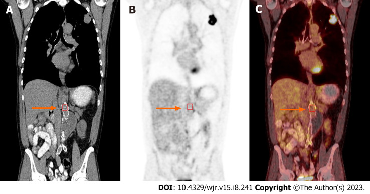
Diagnosis of prosthetic vascular graft infection with [(18)F]fluorodeoxyglucose positron emission tomography/computed tomography (18F-FDG PET/CT) allows for early detection of functional changes associated with infection, based on increased glucose utilization by activated macrophages and granulocytes. Aseptic vascular grafts, like all foreign bodies, can stimulate an inflammatory response, which can present as increased activity on 18F-FDG PET/CT. Consequently, distinguishing aseptic inflammation from graft infection, though important, can be difficult. In the case of endovascular aneurysm repair (EVAR), a minimally invasive procedure involving the transfemoral insertion of an endoprosthetic stent graft, the normal postoperative appearance of these grafts on 18F-FDG PET/CT can vary over time, potentially confounding study interpretation.

AIM

To investigate the visual, semiquantitative, and temporal characteristics of aseptic vascular grafts in patients status post EVAR.

METHODS

In this observational retrospective cohort study, patients with history of EVAR who underwent 18F-FDG PET/CT for indications other than infection were identified retrospectively. All patients were asymptomatic for graft infection - no abdominal pain, fever of unknown origin, sepsis, or leukocytosis - at the time of imaging and for ≥ 2 mo after each PET/CT. Imaging studies such as CT for each patient were also reviewed, and any patients with suspected or confirmed vascular graft infection were excluded. One hundred two scans performed on 43 patients (34 males; 9 females; age = 77 ± 8 years at the time of the final PET/CT) were retrospectively reviewed. All 43 patients had an abdominal aortic (AA) vascular graft, 40 patients had a right iliac (RI) limb graft, and 41 patients had a left iliac (LI) limb graft. Twenty-two patients had 1 PET/CT and 21 patients had from 2 to 9 PET/CTs. Grafts were imaged between 2 mo to 168 mo (about 14 years) post placement. Eight grafts were imaged within 6 mo of placement, including three that were imaged within three months of placement. The mean interval between graft placement and PET/CT for all 102 scans was 51 ± 39 mo. PET/CT data was reconstructed with region-of-interest analysis of proximal, mid and distal portions of the grafts and background ascending aorta. Maximum standardized uptake value (SUV) was recorded for each region. SUV-to-background uptake ratios (URs) were calculated. Visual assessment was performed using a 2-pattern grading scale: Diffuse (homogeneous uptake less than liver uptake) and focal (one or more areas of focal uptake in any part of the graft). Statistical analysis was performed.

RESULTS

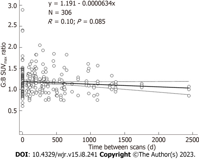
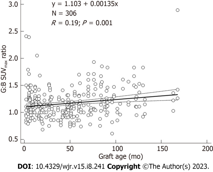
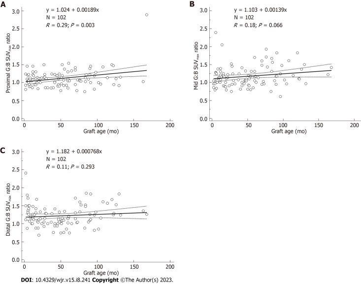
In total, there were 306 AA grafts, 285 LI grafts, 282 RI grafts, and 306 ascending aorta background SUV measurements. For all 102 scans, mean SUV values for AA grafts were 2.8-3.0 along proximal, mid, and distal segments. Mean SUV values for LI grafts and RI grafts were 2.7-2.8. Mean SUV values for background were 2.5 ± 0.5. Mean URs were 1.1-1.2. Visual analysis of the scans reflected results of quantitative analysis. On visual inspection, 98% revealed diffuse, homogeneous 18F-FDG uptake less than liver. Graft URs and visual pattern categories were significantly associated for AA graft URs (F-ratio = 21.5, 0.001), LI graft URs (F-ratio = 20.4, 0.001), and RI graft URs (F-ratio = 30.4, 0.001). Thus, visual patterns of 18F-FDG uptake corresponded statistically significantly to semiquantitative URs. The age of grafts showing focal patterns was greater than grafts showing diffuse patterns, 87 ± 89 50 ± 37 mo, respectively ( = 0.02). URs were significantly associated with graft age for AA grafts ( = 0.19, = 0.001). URs were also significantly associated with graft age for LI grafts ( = 0.25, < 0.0001), and RI grafts ( = 0.31, < 0.001). Quartiles of similar numbers of graft ( = 25-27) grouped by graft age indicated that URs were significantly higher for 4 quartile 2 quartile URs (F-ratio = 19.5, 0.001). When evaluating URs, graft SUVmax values within 10%-20% of the ascending aorta SUV is evident in aseptic grafts, except for grafts in the oldest quartiles. In this study, grafts in the oldest quartiles (> 7 years post EVAR) showed SUV up to 30% higher than the ascending aorta SUV.

CONCLUSION

Characteristics of an aseptic vascular stent graft in the aorta and iliac vessels on 18F-FDG PET/CT include graft SUV values within 10%-20% of the ascending aorta background SUV. The SUV of older aseptic grafts can be as much as 30% above background. The visual uptake pattern of diffuse, homogeneous uptake less than liver was seen in 98% of aseptic vascular grafts, making this pattern particularly reassuring for clinicians.

摘要

背景

使用[¹⁸F]氟脱氧葡萄糖正电子发射断层扫描/计算机断层扫描(¹⁸F-FDG PET/CT)诊断人工血管移植物感染,基于活化巨噬细胞和粒细胞对葡萄糖利用增加,能够早期检测与感染相关的功能变化。无菌血管移植物与所有异物一样,可刺激炎症反应,在¹⁸F-FDG PET/CT上可表现为活性增加。因此,区分无菌性炎症与移植物感染虽然很重要,但可能具有挑战性。在血管内动脉瘤修复术(EVAR)中,这是一种涉及经股动脉插入腔内人工血管支架的微创手术,这些移植物在¹⁸F-FDG PET/CT上的正常术后表现会随时间变化,这可能会混淆研究结果的解读。

目的

研究接受EVAR术后患者的无菌性血管移植物的视觉、半定量和时间特征。

方法

在这项观察性回顾性队列研究中,对有EVAR病史且因非感染指征接受¹⁸F-FDG PET/CT检查的患者进行回顾性识别。所有患者在成像时以及每次PET/CT检查后≥2个月均无移植物感染症状——无腹痛、不明原因发热、败血症或白细胞增多。还对每位患者的CT等影像学检查进行了回顾,并排除了任何疑似或确诊血管移植物感染的患者。对43例患者(34例男性;9例女性;最后一次PET/CT检查时年龄为77±8岁)进行的102次扫描进行了回顾性分析。所有43例患者均有腹主动脉(AA)血管移植物,40例患者有右髂(RI)肢体移植物,41例患者有左髂(LI)肢体移植物。22例患者进行了1次PET/CT检查,21例患者进行了2至9次PET/CT检查。移植物在植入后2个月至168个月(约14年)进行成像。8例移植物在植入后6个月内成像,其中3例在植入后3个月内成像。所有102次扫描中移植物植入与PET/CT检查的平均间隔时间为51±39个月。PET/CT数据通过对移植物近端、中段和远端以及背景升主动脉进行感兴趣区分析进行重建。记录每个区域的最大标准化摄取值(SUV)。计算SUV与背景摄取比值(UR)。使用2种模式分级量表进行视觉评估:弥漫性(均匀摄取低于肝脏摄取)和局灶性(移植物任何部位有一个或多个局灶性摄取区域)。进行了统计分析。

结果

总共对306个AA移植物、285个LI移植物、282个RI移植物以及306个升主动脉背景SUV测量值进行了分析。在所有102次扫描中,AA移植物近端、中段和远端节段的平均SUV值为2.8 - 3.0。LI移植物和RI移植物的平均SUV值为2.7 - 2.8。背景的平均SUV值为2.5±0.5。平均UR为1.1 - 1.2。扫描的视觉分析反映了定量分析结果。在视觉检查中,98%显示弥漫性、均匀的¹⁸F-FDG摄取低于肝脏。AA移植物UR(F值 = 21.5,P = 0.001)、LI移植物UR(F值 = 20.4,P = 0.001)和RI移植物UR(F值 = 30.4,P = 0.001)的移植物UR与视觉模式类别显著相关。因此,¹⁸F-FDG摄取的视觉模式在统计学上与半定量UR显著对应。显示局灶性模式的移植物年龄大于显示弥漫性模式的移植物,分别为87±89个月和50±37个月(P = 0.02)。AA移植物的UR与移植物年龄显著相关(r = 0.19,P = 0.001)。LI移植物的UR也与移植物年龄显著相关(r = 0.25,P < 0.0001),RI移植物的UR与移植物年龄显著相关(r = 0.31,P < 0.001)。按移植物年龄分组的相似数量移植物(n = 25 - 27)四分位数表明,第4四分位数的UR显著高于第2四分位数的UR(F值 = 19.5,P = 0.001)。在评估UR时,除了最老四分位数的移植物外,无菌移植物中移植物SUVmax值在升主动脉SUV的10% - 20%范围内是明显的。在本研究中,最老四分位数(EVAR术后>7年)的移植物显示SUV比升主动脉SUV高30%。

结论

¹⁸F-FDG PET/CT上主动脉和髂血管中无菌性血管支架移植物的特征包括移植物SUV值在升主动脉背景SUV的10% - 20%范围内。较老无菌移植物的SUV可比背景高30%。98%的无菌血管移植物可见弥漫性、均匀摄取低于肝脏的视觉摄取模式,这一模式对临床医生特别有参考价值。

https://cdn.ncbi.nlm.nih.gov/pmc/blobs/83b3/10473915/54b290990065/WJR-15-241-g004.jpg
https://cdn.ncbi.nlm.nih.gov/pmc/blobs/83b3/10473915/5e1436537b1f/WJR-15-241-g001.jpg
https://cdn.ncbi.nlm.nih.gov/pmc/blobs/83b3/10473915/2585be27c74b/WJR-15-241-g002.jpg
https://cdn.ncbi.nlm.nih.gov/pmc/blobs/83b3/10473915/3617e2b057a9/WJR-15-241-g003.jpg
https://cdn.ncbi.nlm.nih.gov/pmc/blobs/83b3/10473915/54b290990065/WJR-15-241-g004.jpg

相似文献

1
Appearance of aseptic vascular grafts after endovascular aortic repair on [(18)F]fluorodeoxyglucose positron emission tomography/computed tomography.经皮腔内主动脉修复术后无菌血管移植物在[(18)F]氟脱氧葡萄糖正电子发射断层扫描/计算机断层扫描上的表现
World J Radiol. 2023 Aug 28;15(8):241-249. doi: 10.4329/wjr.v15.i8.241.
2
Determining the Diagnostic Value of 18F-Fluorodeoxyglucose Positron Emission/Computed Tomography in Detecting Prosthetic Aortic Graft Infection.确定18F-氟脱氧葡萄糖正电子发射断层扫描/计算机断层扫描在检测人工主动脉移植物感染中的诊断价值。
Ann Vasc Surg. 2018 Nov;53:78-85. doi: 10.1016/j.avsg.2018.04.028. Epub 2018 Aug 11.
3
More advantages in detecting bone and soft tissue metastases from prostate cancer using F-PSMA PET/CT.使用F-PSMA PET/CT检测前列腺癌骨和软组织转移方面有更多优势。
Hell J Nucl Med. 2019 Jan-Apr;22(1):6-9. doi: 10.1967/s002449910952. Epub 2019 Mar 7.
4
The relevance of 18F-fluorodeoxyglucose positron emission tomography/computed tomography imaging in diagnosing prosthetic graft infections post cardiac and proximal thoracic aortic surgery.18F-氟脱氧葡萄糖正电子发射断层扫描/计算机断层扫描成像在诊断心脏及胸主动脉近端手术后人工血管感染中的相关性
Interact Cardiovasc Thorac Surg. 2015 Oct;21(4):450-8. doi: 10.1093/icvts/ivv178. Epub 2015 Jul 13.
5
[F]FDG Uptake in Non-Infected Endovascular Grafts: A Retrospective Study.[F]非感染性血管内移植物中的氟代脱氧葡萄糖摄取:一项回顾性研究。
Diagnostics (Basel). 2023 Jan 23;13(3):409. doi: 10.3390/diagnostics13030409.
6
Detection of thoracic aortic prosthetic graft infection with 18F-fluorodeoxyglucose positron emission tomography/computed tomography.18F-氟脱氧葡萄糖正电子发射断层扫描/计算机断层扫描检测胸主动脉人工移植物感染。
Eur J Cardiothorac Surg. 2013 Jun;43(6):1183-7. doi: 10.1093/ejcts/ezs693. Epub 2013 Jan 18.
7
18F-fluorodeoxyglucose uptake patterns in positron emission tomography/computed tomography caused by inflammation and/or infection after graft surgery for thoracic aortic dissection.移植手术后胸主动脉夹层炎症和/或感染引起的正电子发射断层扫描/计算机断层扫描 18F-氟脱氧葡萄糖摄取模式。
J Nucl Cardiol. 2024 Jun;36:101865. doi: 10.1016/j.nuclcard.2024.101865. Epub 2024 Apr 27.
8
18-Fluorodeoxyglucose positron emission tomography in the diagnosis of prosthetic aortic graft infection: the difference between open and endovascular repair.18-氟脱氧葡萄糖正电子发射断层扫描在人工主动脉移植物感染诊断中的应用:开放手术与血管腔内修复的差异
Eur J Cardiothorac Surg. 2022 Dec 2;63(1). doi: 10.1093/ejcts/ezac542.
9
How to recognize stent graft infection after endovascular aortic repair: the utility of 18F-FDG PET/CT in an infrequent but serious clinical setting.如何在血管内主动脉修复后识别支架移植物感染:18F-FDG PET/CT 在罕见但严重的临床情况下的应用。
Ann Nucl Med. 2019 Aug;33(8):594-605. doi: 10.1007/s12149-019-01370-9. Epub 2019 May 29.
10
Low baseline and subsequent higher aortic abdominal aneurysm FDG uptake are associated with poor sac shrinkage post endovascular repair.基线水平低且随后腹主动脉瘤 FDG 摄取较高与血管内修复后瘤囊缩小不良有关。
Eur J Nucl Med Mol Imaging. 2018 Apr;45(4):549-557. doi: 10.1007/s00259-017-3883-1. Epub 2017 Nov 21.

引用本文的文献

1
Knowledge mapping of macrophage and its role in aneurysm from 1999 to 2022: A bibliometric analysis.1999年至2022年巨噬细胞及其在动脉瘤中作用的知识图谱:一项文献计量分析
Heliyon. 2024 Jul 30;10(15):e35212. doi: 10.1016/j.heliyon.2024.e35212. eCollection 2024 Aug 15.

本文引用的文献

1
18-Fluorodeoxyglucose positron emission tomography in the diagnosis of prosthetic aortic graft infection: the difference between open and endovascular repair.18-氟脱氧葡萄糖正电子发射断层扫描在人工主动脉移植物感染诊断中的应用:开放手术与血管腔内修复的差异
Eur J Cardiothorac Surg. 2022 Dec 2;63(1). doi: 10.1093/ejcts/ezac542.
2
FDG PET/CT in abdominal aortic graft infection: A case report and literature review.氟代脱氧葡萄糖正电子发射断层显像/计算机断层扫描在腹主动脉移植物感染中的应用:一例病例报告及文献综述
Radiol Case Rep. 2022 Oct 27;18(1):27-30. doi: 10.1016/j.radcr.2022.09.106. eCollection 2023 Jan.
3
Vascular Graft Infection Imaging.
血管移植物感染的影像学检查。
Semin Nucl Med. 2023 Jan;53(1):70-77. doi: 10.1053/j.semnuclmed.2022.08.006. Epub 2022 Sep 11.
4
Diagnostic Accuracy of FDG PET for the Identification of Vascular Graft Infection.18F-FDG PET 对血管移植物感染的诊断准确性。
Ann Vasc Surg. 2022 Nov;87:422-429. doi: 10.1016/j.avsg.2022.05.029. Epub 2022 Jun 25.
5
Prosthetic vascular graft infection: A systematic review and meta-analysis on diagnostic accuracy of 18FDG PET/CT.人工血管移植物感染:18FDG PET/CT 诊断准确性的系统评价和荟萃分析。
Gen Thorac Cardiovasc Surg. 2022 Mar;70(3):219-229. doi: 10.1007/s11748-021-01682-6. Epub 2021 Jul 26.
6
State of the art of F-FDG PET/CT application in inflammation and infection: a guide for image acquisition and interpretation.F-FDG PET/CT在炎症和感染中的应用现状:图像采集与解读指南
Clin Transl Imaging. 2021;9(4):299-339. doi: 10.1007/s40336-021-00445-w. Epub 2021 Jul 10.
7
Patterns of vascular graft infection in 18F-FDG PET/CT.18F-FDG PET/CT 中的血管移植物感染模式。
Nucl Med Rev Cent East Eur. 2020;23(2):63-70. doi: 10.5603/NMR.a2020.0015.
8
Vascular findings on FDG PET/CT.FDG PET/CT 中的血管所见。
Br J Radiol. 2020 Sep 1;93(1113):20200103. doi: 10.1259/bjr.20200103. Epub 2020 May 6.
9
18F-FDG PET in the Diagnosis of Vascular Prosthetic Graft Infection: A Diagnostic Test Accuracy Meta-Analysis.18F-FDG PET 在血管移植物感染诊断中的应用:一项诊断试验准确性的荟萃分析。
Eur J Vasc Endovasc Surg. 2019 Feb;57(2):292-301. doi: 10.1016/j.ejvs.2018.08.040. Epub 2018 Sep 18.
10
Outcomes of Surgical Explantation of Infected Aortic Grafts After Endovascular and Open Abdominal Aneurysm Repair.血管内和开放腹主动脉瘤修复术后感染性主动脉移植物的外科取出的结果。
Eur J Vasc Endovasc Surg. 2019 Jan;57(1):130-136. doi: 10.1016/j.ejvs.2018.07.021. Epub 2018 Aug 23.