• 文献检索
  • 文档翻译
  • 深度研究
  • 学术资讯
  • Suppr Zotero 插件Zotero 插件
  • 邀请有礼
  • 套餐&价格
  • 历史记录
应用&插件
Suppr Zotero 插件Zotero 插件浏览器插件Mac 客户端Windows 客户端微信小程序
定价
高级版会员购买积分包购买API积分包
服务
文献检索文档翻译深度研究API 文档MCP 服务
关于我们
关于 Suppr公司介绍联系我们用户协议隐私条款
关注我们

Suppr 超能文献

核心技术专利:CN118964589B侵权必究
粤ICP备2023148730 号-1Suppr @ 2026

文献检索

告别复杂PubMed语法,用中文像聊天一样搜索,搜遍4000万医学文献。AI智能推荐,让科研检索更轻松。

立即免费搜索

文件翻译

保留排版,准确专业,支持PDF/Word/PPT等文件格式,支持 12+语言互译。

免费翻译文档

深度研究

AI帮你快速写综述,25分钟生成高质量综述,智能提取关键信息,辅助科研写作。

立即免费体验

新冠病毒感染后综合征中小气道疾病,长新冠患者三年随访中的一种非传统方法:病例报告。

Small airway disease in post-acute COVID-19 syndrome, a non-conventional approach in three years follow-up of a patient with long COVID: a case report.

机构信息

Universidad Espíritu Santo, Km. 2.5 Vía La Puntilla, Samborondón, 0901-952, Ecuador.

Respiralab, Respiralab Research Group, Guayaquil, Ecuador.

出版信息

J Med Case Rep. 2023 Sep 11;17(1):386. doi: 10.1186/s13256-023-04113-7.

DOI:10.1186/s13256-023-04113-7
PMID:37691104
原文链接:https://pmc.ncbi.nlm.nih.gov/articles/PMC10494419/
Abstract

BACKGROUND

Small airways disease (SAD), a novel finding described in post-acute COVID-19 patients, should be suspected when respiratory symptoms continue, air trapping persists on expiratory CT scans, and imaging findings fail to improve despite objectively better conventional pulmonary function test (PFT) parameters. The forced oscillation technique (FOT) and Multiple breathing washout (MBW) are both very sensitive methods for detecting anomalies in the peripheral airways.

CASE PRESENTATION

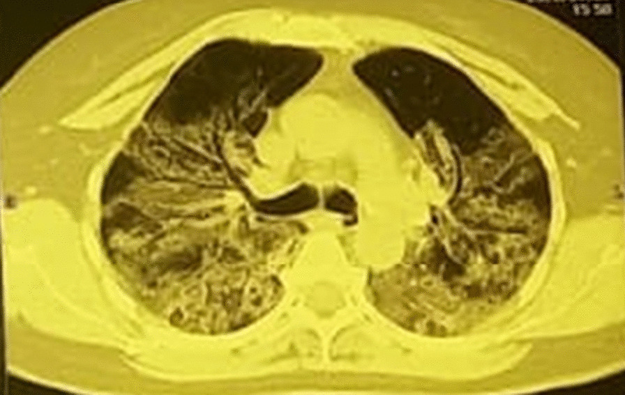
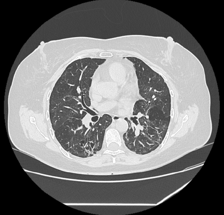
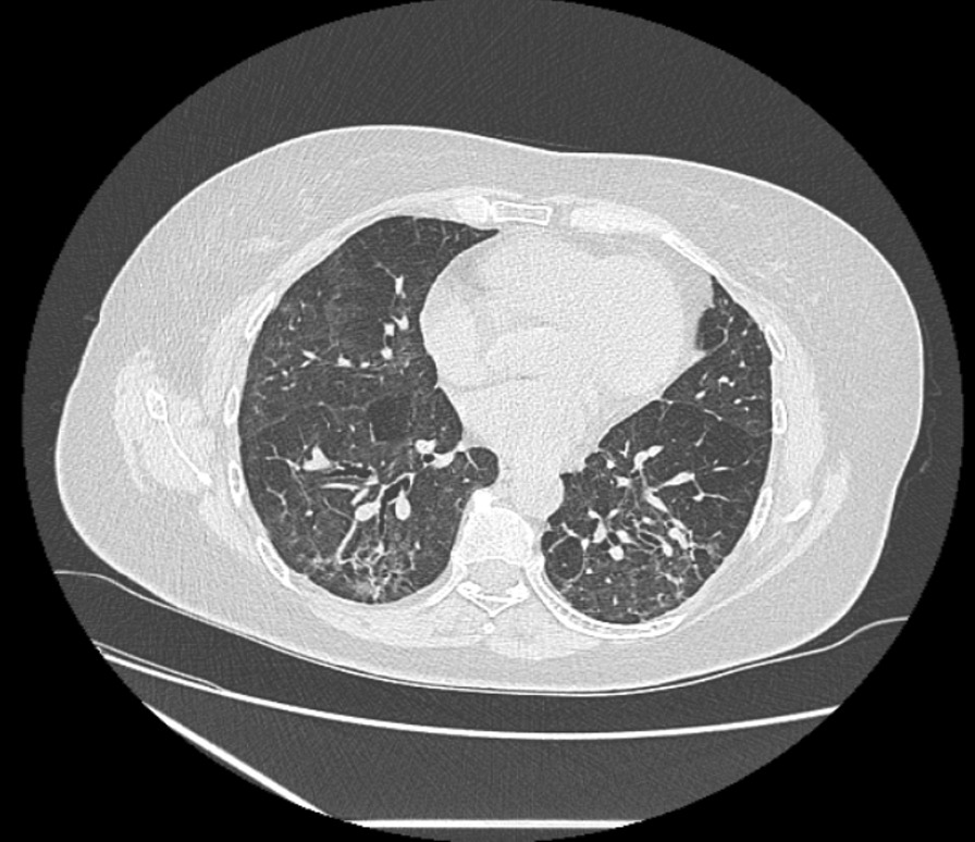
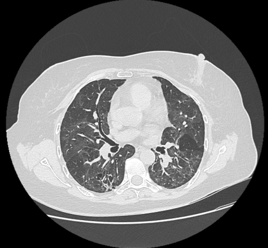
We discuss the case of a 60-year-old Hispanic patient who had severe COVID-19 pneumonia and developed dyspnea, fatigue, and limited daily activity a year later. The PFTs revealed restrictive lung disease, as seen by significant diffusing capacity of the lungs for carbon monoxide (DLCO) decrease, severe desaturation, and poor 6-min walk test (6MWT) performance. The patient was treated with lowering corticosteroids as well as pulmonary rehabilitation (PR). During the 24-month follow-up, the dyspnea and fatigue persisted. On PFTs, 6MWT performance and restricted pattern improved slightly, but MBW discovered significant ventilatory inhomogeneity. FOT revealed substantial peripheral airway obstructive abnormalities. On CT scans, air trapping and ground-glass opacities (GGO) improved somewhat. The patient used a bronchodilator twice a day and low-dose inhaled corticosteroids (160 µg of budesonide and 4.5 µg of formoterol fumarate dihydrate) for nine months. PR sessions were resuming. The restricting parameters were stabilized and the DLCO had normalized after 36 months, with a 6MWT performance of 87% but significant desaturation. The CT scan revealed traction bronchiectasis, low GGO, and persistent air trapping. Without normalization, FOT and MBW scores improved, indicating small airway disease.

CONCLUSIONS

The necessity of integrating these tests when detecting SAD is emphasized in our paper. This article lays the foundation for future research into the best ways to manage and monitor SAD in post-acute COVID-19 patients.

摘要

背景

小气道疾病(SAD)是一种在急性 COVID-19 后患者中描述的新发现,如果出现以下情况,应怀疑存在 SAD:呼吸症状持续存在、呼气 CT 扫描显示空气滞留、尽管常规肺功能测试(PFT)参数客观改善,但影像学结果无改善。强迫振荡技术(FOT)和多次呼吸清除(MBW)都是检测外周气道异常的非常敏感的方法。

病例介绍

我们讨论了一位 60 岁的西班牙裔患者的病例,他患有严重的 COVID-19 肺炎,一年后出现呼吸困难、疲劳和日常活动受限。PFT 显示为限制性肺病,表现为一氧化碳弥散量(DLCO)显著降低、严重低氧血症和 6 分钟步行测试(6MWT)表现不佳。患者接受了降低皮质激素和肺康复(PR)治疗。在 24 个月的随访中,呼吸困难和疲劳持续存在。在 PFT 上,6MWT 表现和限制性模式略有改善,但 MBW 发现显著的通气不均一性。FOT 显示出显著的外周气道阻塞异常。在 CT 扫描上,空气滞留和磨玻璃影(GGO)有所改善。患者每天使用支气管扩张剂两次,并使用低剂量吸入皮质激素(布地奈德 160µg 和富马酸福莫特罗二水合物 4.5µg)治疗 9 个月。PR 课程正在恢复。限制参数稳定,DLCO 在 36 个月后恢复正常,6MWT 表现为 87%,但仍存在显著低氧血症。CT 扫描显示牵引性支气管扩张、低 GGO 和持续的空气滞留。尽管未恢复正常,但 FOT 和 MBW 评分有所改善,表明存在小气道疾病。

结论

本文强调了在检测 SAD 时整合这些测试的必要性。本文为研究急性 COVID-19 后患者的 SAD 最佳管理和监测方法奠定了基础。

https://cdn.ncbi.nlm.nih.gov/pmc/blobs/a06a/10494419/395add35dce3/13256_2023_4113_Fig4_HTML.jpg
https://cdn.ncbi.nlm.nih.gov/pmc/blobs/a06a/10494419/508b2d0da36b/13256_2023_4113_Fig1_HTML.jpg
https://cdn.ncbi.nlm.nih.gov/pmc/blobs/a06a/10494419/834cd56c0fda/13256_2023_4113_Fig2_HTML.jpg
https://cdn.ncbi.nlm.nih.gov/pmc/blobs/a06a/10494419/e8d3bd67e706/13256_2023_4113_Fig3_HTML.jpg
https://cdn.ncbi.nlm.nih.gov/pmc/blobs/a06a/10494419/395add35dce3/13256_2023_4113_Fig4_HTML.jpg

相似文献

1
Small airway disease in post-acute COVID-19 syndrome, a non-conventional approach in three years follow-up of a patient with long COVID: a case report.新冠病毒感染后综合征中小气道疾病,长新冠患者三年随访中的一种非传统方法:病例报告。
J Med Case Rep. 2023 Sep 11;17(1):386. doi: 10.1186/s13256-023-04113-7.
2
Forced Oscillation Technique and Small Airway Involvement in Chronic Hypersensitivity Pneumonitis.强迫振荡技术与慢性过敏性肺炎中的小气道受累
Arch Bronconeumol (Engl Ed). 2019 Oct;55(10):519-525. doi: 10.1016/j.arbres.2019.01.022. Epub 2019 Apr 8.
3
Long-term dyspnea, regional ventilation distribution and peripheral lung function in COVID-19 survivors: a 1 year follow up study.COVID-19 幸存者的长期呼吸困难、区域性通气分布和肺外周功能:1 年随访研究。
BMC Pulm Med. 2022 Nov 9;22(1):408. doi: 10.1186/s12890-022-02214-5.
4
A Follow-Up Study of Lung Function and Chest Computed Tomography at 6 Months after Discharge in Patients with Coronavirus Disease 2019.新冠肺炎患者出院后 6 个月时的肺功能和胸部计算机断层扫描随访研究。
Can Respir J. 2021 Feb 13;2021:6692409. doi: 10.1155/2021/6692409. eCollection 2021.
5
[Chinese experts consensus statement: diagnosis and treatment of cystic fibrosis (2023)].[中国专家共识声明:囊性纤维化的诊断与治疗(2023年)]
Zhonghua Jie He He Hu Xi Za Zhi. 2023 Apr 12;46(4):352-372. doi: 10.3760/cma.j.cn112147-20221214-00971.
6
Impaired function in the lung periphery following COVID-19 is associated with lingering breathing difficulties.新冠肺炎后肺外周功能受损与持续存在的呼吸困难有关。
Physiol Rep. 2024 Jan;12(2):e15918. doi: 10.14814/phy2.15918.
7
Air trapping in COVID-19 patients following hospital discharge: retrospective evaluation with paired inspiratory/expiratory thin-section CT.COVID-19 患者出院后患气腔滞留:采用配对吸气/呼气薄层 CT 的回顾性评估。
Eur Radiol. 2022 Jul;32(7):4427-4436. doi: 10.1007/s00330-022-08580-2. Epub 2022 Feb 28.
8
Long COVID symptoms after 8-month recovery: persistent static lung hyperinflation associated with small airway dysfunction.8 个月康复后出现的长期新冠症状:持续性静态肺过度充气与小气道功能障碍相关。
Respir Res. 2024 May 15;25(1):209. doi: 10.1186/s12931-024-02830-1.
9
Personalized pulmonary rehabilitation program for patients with post-acute sequelae of COVID-19: A proof-of-concept retrospective study.针对 COVID-19 后急性后遗症患者的个性化肺康复计划:一项概念验证回顾性研究。
Physiol Rep. 2024 Feb;12(3):e15931. doi: 10.14814/phy2.15931.
10
Pulmonary function tests in the follow-up of children with COVID-19.儿童 COVID-19 随访中的肺功能检查。
Eur J Pediatr. 2022 Jul;181(7):2839-2847. doi: 10.1007/s00431-022-04493-w. Epub 2022 May 6.

引用本文的文献

1
New-onset obstructive airway disease following COVID-19: a multicenter retrospective cohort study.新型冠状病毒病后新发阻塞性气道疾病:一项多中心回顾性队列研究。
BMC Med. 2024 Sep 4;22(1):360. doi: 10.1186/s12916-024-03589-4.
2
Increased Expression of ATP12A in Small Airway Epithelia of Post-COVID-19 Pulmonary Fibrosis.新冠后肺纤维化患者小气道上皮细胞中ATP12A表达增加
Am J Respir Cell Mol Biol. 2024 Jun;70(6):527-530. doi: 10.1165/rcmb.2023-0419LE.
3
The impact of social distancing measures (quarantine) policy on tertiary education and medical consultations in China during the COVID-19 pandemic.

本文引用的文献

1
Postacute COVID-19 Syndrome: Xe MRI Ventilation Defects and Respiratory Outcomes 1 Year Later.急性新冠后综合征:Xe MRI 通气缺陷与 1 年后的呼吸结局。
Radiology. 2023 Apr;307(2):e222557. doi: 10.1148/radiol.222557. Epub 2023 Feb 7.
2
Relationship between exercise capacity and impulse oscillometry parameters after COVID-19 infections.新冠病毒感染后运动能力与脉冲震荡参数的关系。
Wien Klin Wochenschr. 2023 May;135(9-10):260-265. doi: 10.1007/s00508-022-02137-5. Epub 2022 Dec 30.
3
Post-COVID-19 Symptoms 2 Years After SARS-CoV-2 Infection Among Hospitalized vs Nonhospitalized Patients.
新冠疫情期间社交距离措施(隔离)政策对中国高等教育和医疗咨询的影响。
Front Public Health. 2024 Mar 5;12:1365805. doi: 10.3389/fpubh.2024.1365805. eCollection 2024.
4
Factors associated with small airway obstruction in COVID-19 survivors: A cross-sectional study among health-care providers.新冠病毒疾病康复者中小气道阻塞的相关因素:一项针对医护人员的横断面研究
Narra J. 2023 Dec;3(3):e437. doi: 10.52225/narra.v3i3.437. Epub 2023 Nov 13.
5
Post-COVID changes in lung function 6 months after veno-venous extracorporeal membrane oxygenation: a prospective observational clinical trial.静脉-静脉体外膜肺氧合术后6个月的新冠后肺功能变化:一项前瞻性观察性临床试验
Front Med (Lausanne). 2023 Dec 20;10:1288679. doi: 10.3389/fmed.2023.1288679. eCollection 2023.
SARS-CoV-2 感染后 2 年住院与非住院患者的新冠后症状。
JAMA Netw Open. 2022 Nov 1;5(11):e2242106. doi: 10.1001/jamanetworkopen.2022.42106.
4
Challenges in the Management of Post-COVID-19 Pulmonary Fibrosis for the Latin American Population.拉丁美洲人群新冠后肺纤维化管理中的挑战
J Pers Med. 2022 Aug 27;12(9):1393. doi: 10.3390/jpm12091393.
5
The Importance of Radiological Patterns and Small Airway Disease in Long-Term Follow-Up of Postacute COVID-19: A Preliminary Study.放射学模式和小气道疾病在急性后COVID-19长期随访中的重要性:一项初步研究
Radiol Res Pract. 2022 May 5;2022:7919033. doi: 10.1155/2022/7919033. eCollection 2022.
6
Quantitative Chest CT Assessment of Small Airways Disease in Post-Acute SARS-CoV-2 Infection.定量胸部 CT 评估急性 SARS-CoV-2 感染后小气道疾病。
Radiology. 2022 Jul;304(1):185-192. doi: 10.1148/radiol.212170. Epub 2022 Mar 15.
7
Air trapping in COVID-19 patients following hospital discharge: retrospective evaluation with paired inspiratory/expiratory thin-section CT.COVID-19 患者出院后患气腔滞留:采用配对吸气/呼气薄层 CT 的回顾性评估。
Eur Radiol. 2022 Jul;32(7):4427-4436. doi: 10.1007/s00330-022-08580-2. Epub 2022 Feb 28.
8
Clinical significance and applications of oscillometry.容积描记法的临床意义和应用。
Eur Respir Rev. 2022 Feb 9;31(163). doi: 10.1183/16000617.0208-2021. Print 2022 Mar 31.
9
Current Needs Assessment for Using Lung Clearance Index for Asthma in Clinical Practice.肺清除指数在哮喘临床实践中应用的当前需求评估
Curr Allergy Asthma Rep. 2022 Feb;22(2):13-20. doi: 10.1007/s11882-022-01025-2. Epub 2022 Jan 24.
10
Inhaled Medicines: Past, Present, and Future.吸入药物:过去、现在与未来。
Pharmacol Rev. 2022 Jan;74(1):48-118. doi: 10.1124/pharmrev.120.000108.