Center of Biotherapy, Beijing Hospital, National Center of Gerontology; Institute of Geriatric Medicine, Chinese Academy of Medical Sciences, Graduate School of Peking Union Medical College, Beijing, 100730, People's Republic of China.
Savaid Medical School, University of Chinese Academy of Sciences, Beijing, 100049, People's Republic of China.
J Exp Clin Cancer Res. 2023 Sep 11;42(1):237. doi: 10.1186/s13046-023-02804-z.
Chemotherapeutic agents are used to control tumor proliferation. However, their influence in the pre-metastatic niche of target organs has not been well studied. Oxaliplatin (OXA) is a drug applied in standard treatments of colorectal cancer (CRC), while the direct effect of which on the pre-metastatic microenvironment of the liver remains unclear.
Models of liver metastases were established with luciferase expressing CT26 cells in BALB/c and BALB/c-nude mice. Single-cell RNA Sequencing was performed to examine the immune microenvironment in the liver elicited by OXA. Immunofluorescence and flowcytometry were utilized to confirm the changes in the number of immune cells. LDH, CellTrace CFSE Cell Proliferation and apoptosis assays were conducted to explore the impact of OXA on T cells ex vivo. The correlation between chemotherapy-related lymphopenia and metastases was assessed by meta-analysis.
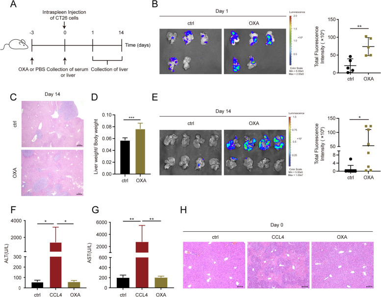
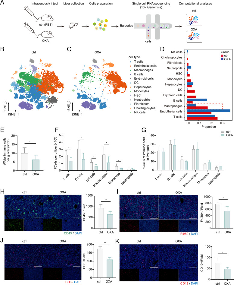
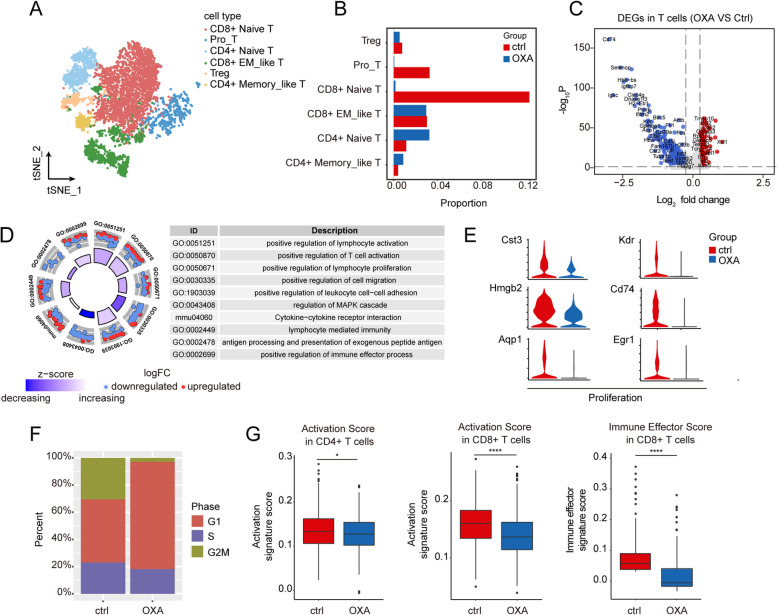
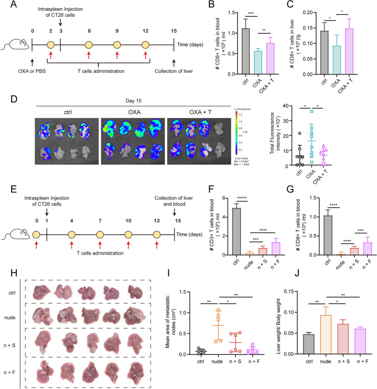
Herein we discovered that administration of OXA prior to the occurrence of liver metastasis actually accelerated tumor development and colonization in the liver. Single-cell RNA sequencing revealed that the landscape of the liver immune microenvironment had been changed to immunosuppressive phenotype. Macrophages after the treatment of OXA exhibited a high ability to inhibit the activation of T cells. Further investigation revealed a significant decrease in the number of T cells in the liver, particularly CD8 T cells with reduced capacity of proliferation, activation, and killing. When mice were treated with T cell supplementation, the OXA-induced metastasis was notably abolished, indicating that the OXA-primed liver microenvironment could be reversed by the infusion of T cells. Consistent with our findings in mice, a meta-analysis was performed to verify that chemotherapy-related lymphopenia was associated with an inferior prognosis related with high incidence of metastasis, suggesting the pivotal role of chemotherapy in pre-metastatic niche formation. Furthermore, a notable reduction in the count of both macrophages and T cells was observed in the liver of colorectal cancer (CRC) patient undergoing OXA-based chemotherapy.
Our findings proposed that immunosuppressive microenvironment in liver induced by OXA enhanced liver metastasis of colorectal cancer, which highlighted a new consideration to balance the pro metastases and anti-cancer possibility of OXA treatment.
化疗药物用于控制肿瘤增殖。然而,它们在靶器官的转移前生态位中的影响尚未得到很好的研究。奥沙利铂(OXA)是一种用于结直肠癌(CRC)标准治疗的药物,但其对肝脏转移前微环境的直接影响尚不清楚。
在 BALB/c 和 BALB/c-nude 小鼠中用表达荧光素酶的 CT26 细胞建立肝转移模型。进行单细胞 RNA 测序以检查 OXA 引起的肝脏免疫微环境中的免疫细胞。免疫荧光和流式细胞术用于确认免疫细胞数量的变化。LDH、CellTrace CFSE 细胞增殖和凋亡测定用于探索 OXA 对体外 T 细胞的影响。通过荟萃分析评估化疗相关淋巴细胞减少与转移之间的相关性。
在此,我们发现,在肝转移发生之前给予 OXA 实际上会加速肿瘤在肝脏中的发展和定植。单细胞 RNA 测序显示,肝脏免疫微环境的景观已发生变化,呈现出免疫抑制表型。经 OXA 处理的巨噬细胞表现出抑制 T 细胞激活的高能力。进一步的研究表明,肝脏中的 T 细胞数量显著减少,特别是增殖、激活和杀伤能力降低的 CD8 T 细胞。当用 T 细胞补充治疗时,OXA 诱导的转移明显被消除,表明 OXA 预激活的肝脏微环境可以通过输注 T 细胞逆转。与我们在小鼠中的发现一致,进行了荟萃分析以验证化疗相关的淋巴细胞减少与高转移发生率相关的预后不良之间的关联,这表明化疗在转移前生态位形成中的关键作用。此外,在接受基于 OXA 的化疗的结直肠癌(CRC)患者的肝脏中观察到巨噬细胞和 T 细胞计数明显减少。
我们的研究结果表明,OXA 诱导的肝脏免疫抑制微环境增强了结直肠癌的肝转移,这突显了在平衡 OXA 治疗的促转移和抗癌可能性方面的新考虑。