Kubota Haruna, Hashimoto Yasushi, Toyota Kazuhiro, Yano Raita, Kobayashi Hironori, Yokoyama Yujiro, Sakashita Yoshihiro, Taniyama Kiyomi, Miyamoto Katsunari, Murakami Yoshiaki
Department of Surgery, Hiroshima Memorial Hospital, Honkawa-cho1-4-3, Naka-ku, Hiroshima, 730-0802, Japan.
Department of Pathology, Hiroshima Memorial Hospital, Hiroshima, Japan.
Surg Case Rep. 2023 Sep 18;9(1):164. doi: 10.1186/s40792-023-01748-y.
Intrahepatic cholangiocarcinoma (ICC) is frequently associated with precursor lesions, and biliary intraepithelial neoplasia (BilIN) may play a significant role in the development of ICC. However, the exact sequence and progression of these lesions remain to be elucidated. We report a rare case of ICC that exhibited extensive longitudinal intraductal extension of high-grade BilIN in the posterior bile ducts and involved the hepatic hilum and the peripheral hepatic parenchyma.
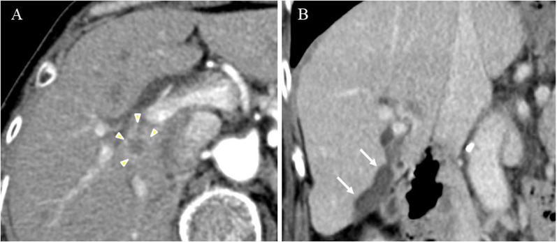
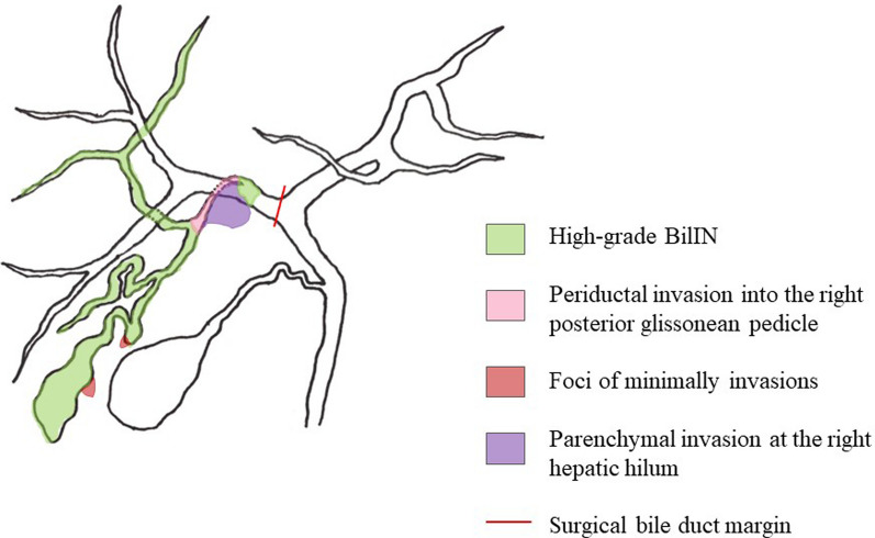
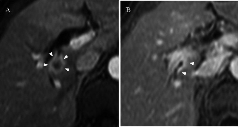
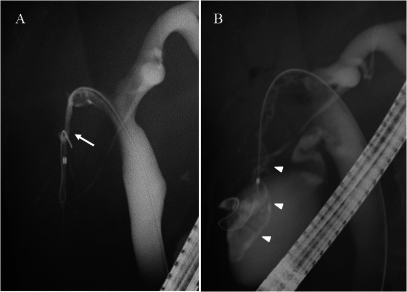
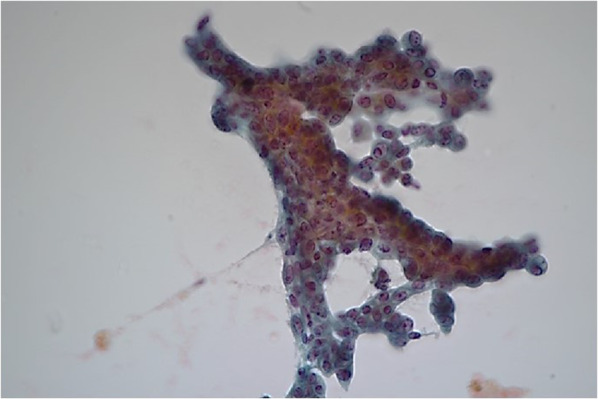
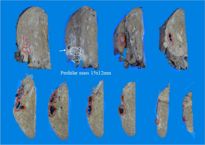
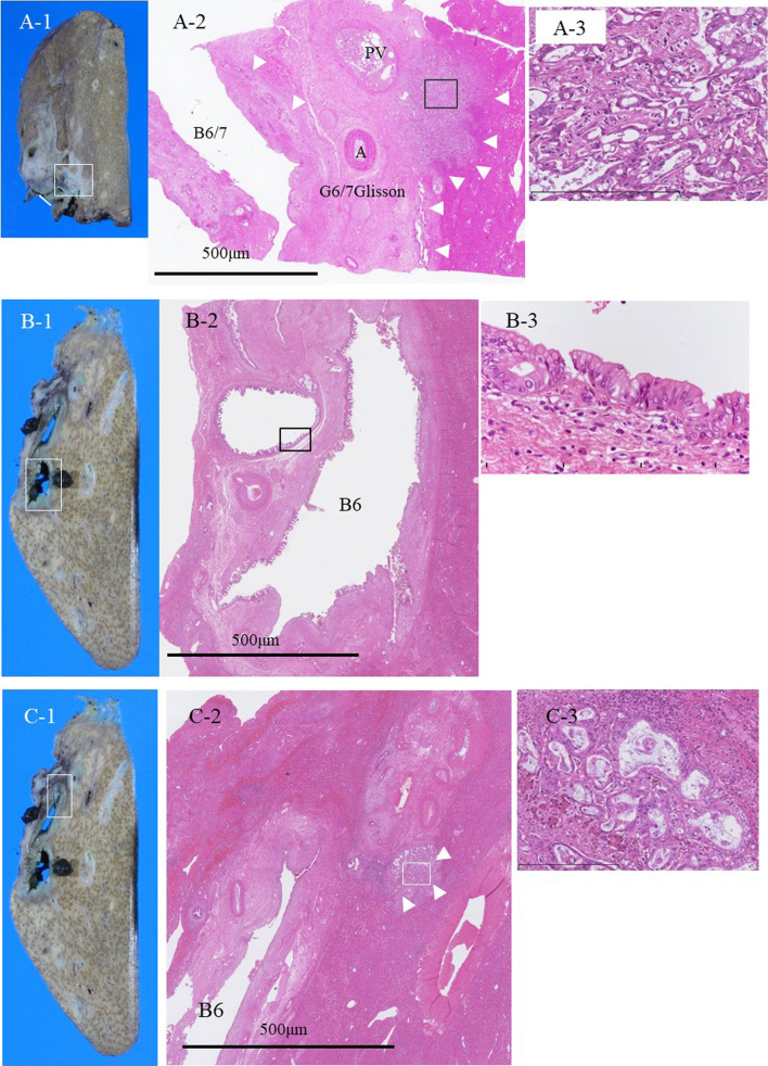
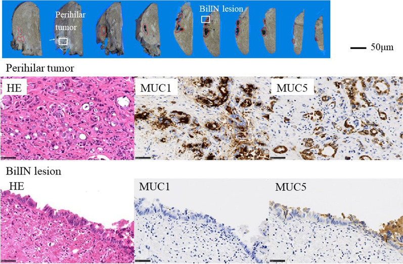
A 70-year-old female presented with anorexia. Computed tomography (CT) revealed a 15 mm enhancing intrahepatic tumor extending to the right intrahepatic secondary confluence. This was associated with a 7 mm diameter cystic dilatation of the segment 6 bile duct (B6). Endoscopic retrograde cholangiopancreatography (ERCP) revealed stenosis at the bifurcation of the posterior bile duct branch. Bile cytology confirmed the diagnosis of adenocarcinoma cells. Therefore, the patient was diagnosed with an ICC involving the right glissonean pedicle and underwent a right hepatectomy and lymph node dissection. Histologic examination revealed the tumor consisted of moderately differentiated adenocarcinoma. In connection with this lesion, diffuse intraductal atypical epithelial cells, which were diagnosed as high-grade BilIN, was observed not only in the dilated B6 but in the entire posterior bile ducts, which measured approximately 120 mm in diameter. Furthermore, two distinct foci of adenocarcinomas were identified in the peripheral hepatic parenchyma. A lymph node metastasis was also present. The pathological diagnosis was ICC pT4N1M0 stage IVA. The patient underwent adjuvant chemotherapy and has shown no recurrence 5 years after surgery. Imaging modalities were unable to accurately assess the extent of the intraductal neoplastic lesions due to their low papillary or sessile intraductal tubular growth. No risk factors for BilIN development, which has the potential to predispose to cholangiocarcinoma, were identified in the present case.
We present a case of ICC involving the right hepatic hilum, accompanied by extensive longitudinal extensions of high-grade BilIN and multifocal microscopic invasions in peripheral hepatic parenchyma. Notably, the intraductal lesions involved the entire posterior intrahepatic bile ducts. The presence of biliary neoplasia with extensive intraductal extension, in conjunction with ICC, should be considered as a variant of BilIN.
肝内胆管癌(ICC)常与癌前病变相关,胆管上皮内瘤变(BilIN)可能在ICC的发生发展中起重要作用。然而,这些病变的确切顺序和进展仍有待阐明。我们报告一例罕见的ICC病例,其在肝后胆管中表现出高级别BilIN广泛的纵向导管内延伸,并累及肝门和肝外周实质。
一名70岁女性因厌食就诊。计算机断层扫描(CT)显示一个15mm强化的肝内肿瘤延伸至肝右叶二级汇合处。这与肝段6胆管(B6)直径7mm的囊性扩张有关。内镜逆行胰胆管造影(ERCP)显示肝后胆管分支分叉处狭窄。胆汁细胞学检查确诊为腺癌细胞。因此,该患者被诊断为累及右肝蒂的ICC,并接受了右肝切除术和淋巴结清扫术。组织学检查显示肿瘤由中分化腺癌组成。与此病变相关的是,不仅在扩张的B6中,而且在整个直径约120mm的肝后胆管中均观察到弥漫性导管内非典型上皮细胞,诊断为高级别BilIN。此外,在肝外周实质中发现了两个不同的腺癌灶。还存在淋巴结转移。病理诊断为ICC pT4N1M0 ⅣA期。该患者接受了辅助化疗,术后5年未复发。由于其乳头状或无蒂导管内管状生长较低,影像学检查无法准确评估导管内肿瘤病变的范围。本病例未发现有发生BilIN的危险因素,而BilIN有可能发展为胆管癌。
我们报告一例累及肝右门的ICC病例,伴有高级别BilIN广泛的纵向延伸和肝外周实质的多灶性微小浸润。值得注意的是,导管内病变累及整个肝后肝内胆管。广泛导管内延伸的胆道肿瘤与ICC并存,应被视为BilIN的一种变异型。