Department of Motor Function Analysis, Human Health Sciences, Graduate School of Medicine, Kyoto University, Kyoto, Japan.
Japan Society for the Promotion of Science, Tokyo, Japan.
PLoS One. 2023 Sep 21;18(9):e0292000. doi: 10.1371/journal.pone.0292000. eCollection 2023.
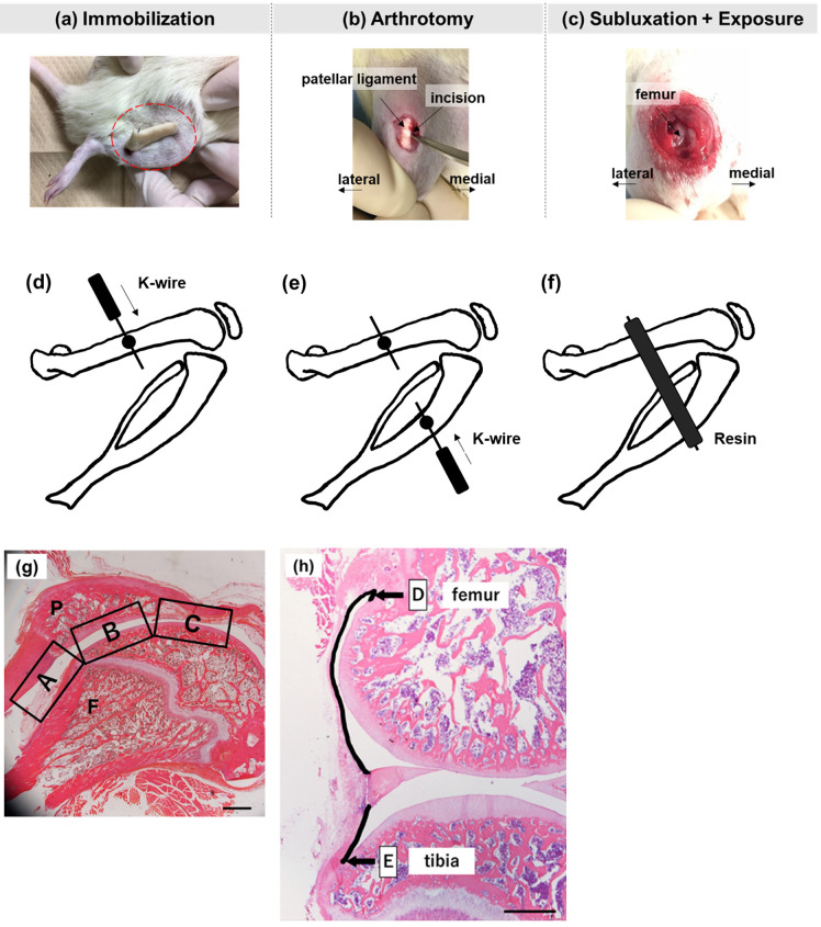
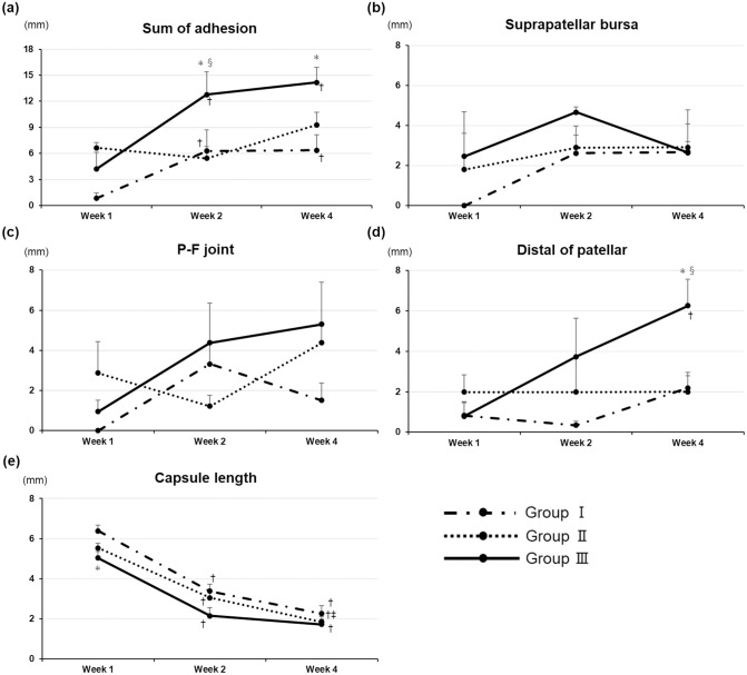
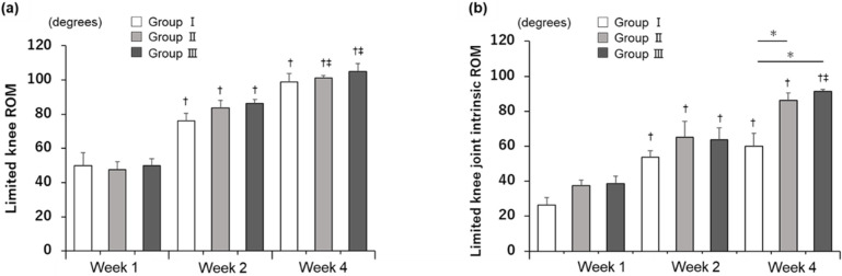
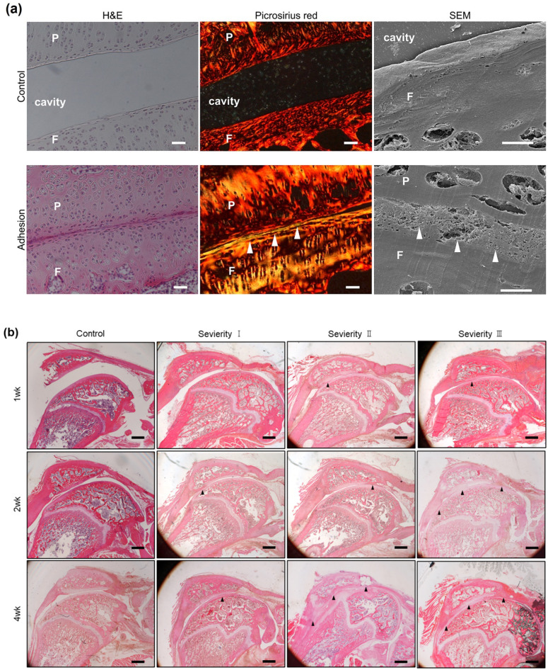
In this study, a novel rat model of knee joint adhesion was developed, and its formation was analyzed quantitatively over time. Thirty-nine Wistar rats were randomly divided into intact control (n = 3) and experimental (n = 36) groups. The latter was equally divided into three groups according to the experimental intervention: fixed with deep bending of the knee joint (group I), fixed after incision of the capsule (group II), and fixed after exposure of the patellofemoral joint to artificial patellar subluxation (group III). All rats were subdivided according to their joint immobilization period (1, 2, or 4 weeks). Thereafter, the limited range of motion of the knee joint with (limited knee range of motion) and without (limited knee joint intrinsic range of motion) skin and muscles were measured. The lengths of adhesions of the anterior knee joint and posterior capsules were evaluated histologically. The limited intrinsic range of motion of the knee joint was found to be increased in groups II and III compared to that in group I 4 weeks after immobilization. Adhesions were confirmed within 1 week after immobilization in groups II and III. The length of the adhesions in group III was significantly longer than in other groups at 2 weeks and remained longer than in group I at 4 weeks. This model may contribute to the assessment of the adhesion process and development of new therapeutic avenues following trauma or surgical invasion.
在这项研究中,建立了一种新型的膝关节粘连大鼠模型,并对其随时间的形成进行了定量分析。39 只 Wistar 大鼠被随机分为完整对照组(n = 3)和实验组(n = 36)。后者根据实验干预分为三组:膝关节深度弯曲固定组(I 组)、囊切开后固定组(II 组)和髌股关节暴露于人工髌骨半脱位后固定组(III 组)。所有大鼠根据关节固定期(1、2 或 4 周)进一步细分。然后,测量膝关节有(膝关节活动度受限)和无(膝关节固有活动度受限)皮肤和肌肉时的活动范围受限程度。对膝关节前侧和后侧囊的粘连长度进行组织学评估。与 I 组相比,固定 4 周后 II 组和 III 组的膝关节固有活动度受限增加。固定后 1 周内 II 组和 III 组可确认粘连。固定 2 周时,III 组的粘连长度明显长于其他组,固定 4 周时仍长于 I 组。该模型可能有助于评估创伤或手术侵袭后的粘连过程,并为新的治疗途径的发展提供依据。