• 文献检索
  • 文档翻译
  • 深度研究
  • 学术资讯
  • Suppr Zotero 插件Zotero 插件
  • 邀请有礼
  • 套餐&价格
  • 历史记录
应用&插件
Suppr Zotero 插件Zotero 插件浏览器插件Mac 客户端Windows 客户端微信小程序
定价
高级版会员购买积分包购买API积分包
服务
文献检索文档翻译深度研究API 文档MCP 服务
关于我们
关于 Suppr公司介绍联系我们用户协议隐私条款
关注我们

Suppr 超能文献

核心技术专利:CN118964589B侵权必究
粤ICP备2023148730 号-1Suppr @ 2026

文献检索

告别复杂PubMed语法,用中文像聊天一样搜索,搜遍4000万医学文献。AI智能推荐,让科研检索更轻松。

立即免费搜索

文件翻译

保留排版,准确专业,支持PDF/Word/PPT等文件格式,支持 12+语言互译。

免费翻译文档

深度研究

AI帮你快速写综述,25分钟生成高质量综述,智能提取关键信息,辅助科研写作。

立即免费体验

高通量数据的使用为巨细胞动脉炎颞动脉过程提供了新的见解。

Use of high-plex data provides novel insights into the temporal artery processes of giant cell arteritis.

机构信息

Division of Rheumatology, Mayo Clinic, Rochester, MN, United States.

Division of Internal Medicine, Dupuytren University Hospital, Limoges, France.

出版信息

Front Immunol. 2023 Sep 6;14:1237986. doi: 10.3389/fimmu.2023.1237986. eCollection 2023.

DOI:10.3389/fimmu.2023.1237986
PMID:37744332
原文链接:https://pmc.ncbi.nlm.nih.gov/articles/PMC10512077/
Abstract

OBJECTIVE

To identify the key coding genes underlying the biomarkers and pathways associated with giant cell arteritis (GCA), we performed an spatial profiling of molecules involved in the temporal arteries of GCA patients and controls. Furthermore, we performed pharmacogenomic network analysis to identify potential treatment targets.

METHODS

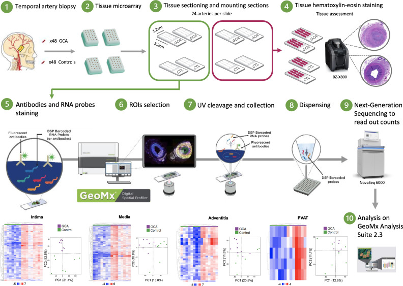
Using human formalin-fixed paraffin-embedded temporal artery biopsy samples (GCA, n = 9; controls, n = 7), we performed a whole transcriptome analysis using the NanoString GeoMx Digital Spatial Profiler. In total, 59 regions of interest were selected in the intima, media, adventitia, and perivascular adipose tissue (PVAT). Differentially expressed genes (DEGs) (fold-change > 2 or < -2, p-adjusted < 0.01) were compared across each layer to build a spatial and pharmacogenomic network and to explore the pathophysiological mechanisms of GCA.

RESULTS

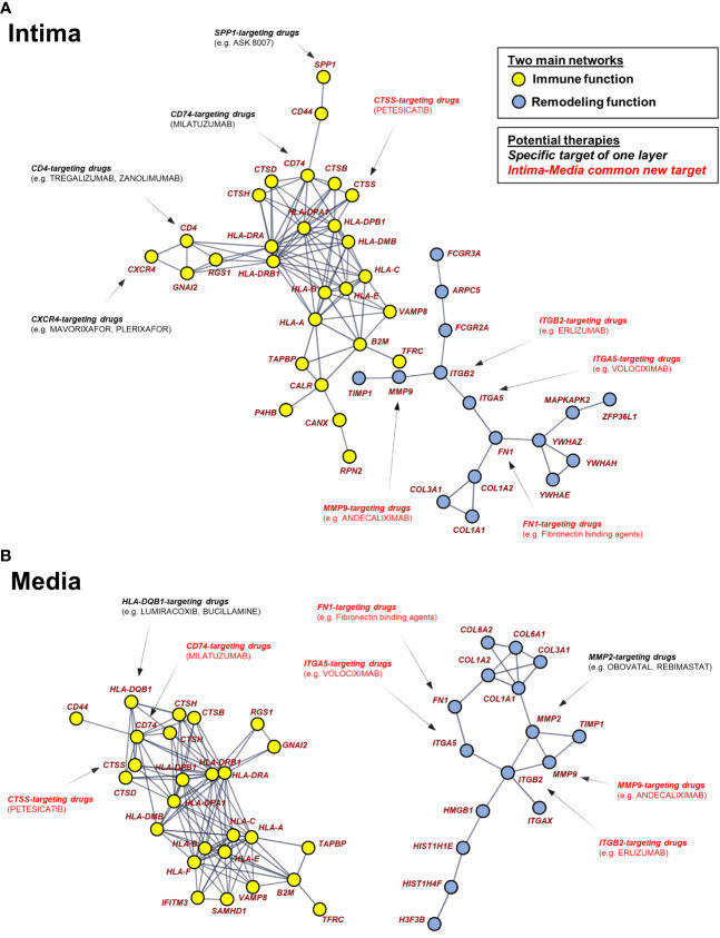
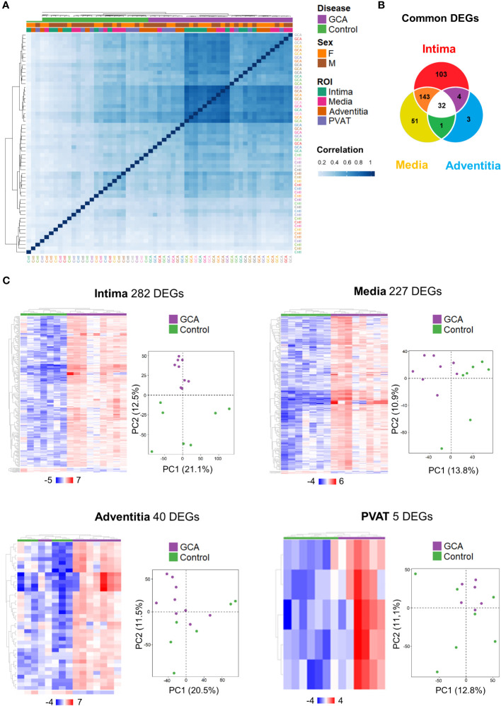
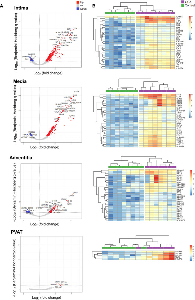
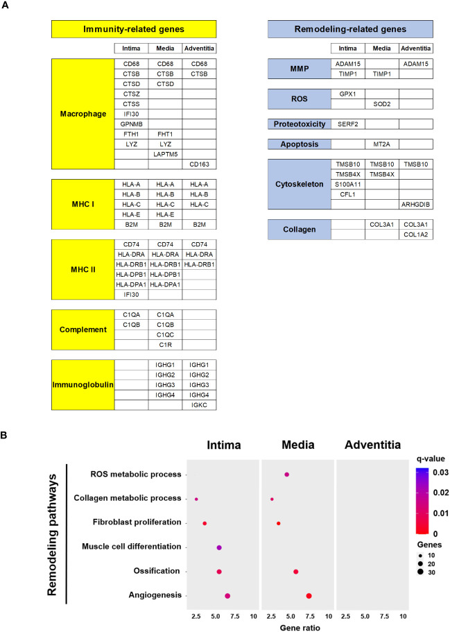
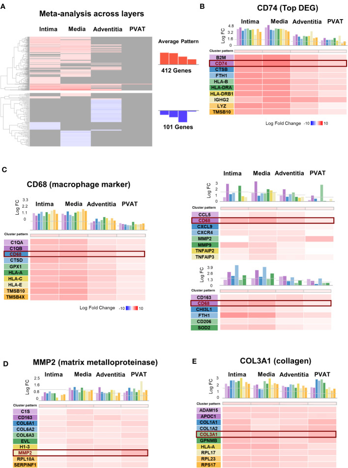
Most of the transcriptome (12,076 genes) was upregulated in GCA arteries, compared to control arteries. Among the screened genes, 282, 227, 40, and 5 DEGs were identified in the intima, media, adventitia, and PVAT, respectively. Genes involved in the immune process and vascular remodeling were upregulated within GCA temporal arteries but differed across the arterial layers. The immune-related functions and vascular remodeling were limited to the intima and media.

CONCLUSION

This study is the first to perform an spatial profiling characterization of the molecules involved in GCA. The pharmacogenomic network analysis identified potential target genes for approved and novel immunotherapies.

摘要

目的

为了确定与巨细胞动脉炎(GCA)相关的生物标志物和途径的关键编码基因,我们对 GCA 患者和对照者的颞动脉中涉及的分子进行了空间分析。此外,我们还进行了药物基因组网络分析,以确定潜在的治疗靶点。

方法

使用福尔马林固定石蜡包埋的人类颞动脉活检样本(GCA,n=9;对照,n=7),我们使用 NanoString GeoMx Digital Spatial Profiler 进行了全转录组分析。总共在内膜、中膜、外膜和血管周围脂肪组织(PVAT)中选择了 59 个感兴趣区域。在各层之间比较差异表达基因(DEG)(fold-change > 2 或 < -2,p-adjusted < 0.01),以构建空间和药物基因组网络,并探索 GCA 的病理生理机制。

结果

与对照动脉相比,GCA 动脉中的大部分转录组(12076 个基因)上调。在筛选出的基因中,内膜、中膜、外膜和 PVAT 分别鉴定出 282、227、40 和 5 个 DEG。参与免疫过程和血管重塑的基因在 GCA 颞动脉中上调,但在动脉各层之间存在差异。免疫相关功能和血管重塑仅限于内膜和中膜。

结论

本研究首次对 GCA 相关分子进行了空间分析。药物基因组网络分析确定了已批准和新型免疫疗法的潜在靶基因。

https://cdn.ncbi.nlm.nih.gov/pmc/blobs/a606/10512077/44ea0cf5dc2b/fimmu-14-1237986-g006.jpg
https://cdn.ncbi.nlm.nih.gov/pmc/blobs/a606/10512077/eacca88c78be/fimmu-14-1237986-g001.jpg
https://cdn.ncbi.nlm.nih.gov/pmc/blobs/a606/10512077/552365a09ee2/fimmu-14-1237986-g002.jpg
https://cdn.ncbi.nlm.nih.gov/pmc/blobs/a606/10512077/55a257366810/fimmu-14-1237986-g003.jpg
https://cdn.ncbi.nlm.nih.gov/pmc/blobs/a606/10512077/4eb14acf2f27/fimmu-14-1237986-g004.jpg
https://cdn.ncbi.nlm.nih.gov/pmc/blobs/a606/10512077/be7c5db17ccd/fimmu-14-1237986-g005.jpg
https://cdn.ncbi.nlm.nih.gov/pmc/blobs/a606/10512077/44ea0cf5dc2b/fimmu-14-1237986-g006.jpg

相似文献

1
Use of high-plex data provides novel insights into the temporal artery processes of giant cell arteritis.高通量数据的使用为巨细胞动脉炎颞动脉过程提供了新的见解。
Front Immunol. 2023 Sep 6;14:1237986. doi: 10.3389/fimmu.2023.1237986. eCollection 2023.
2
An immunohistochemical analysis of fibroblasts in giant cell arteritis.巨细胞动脉炎成纤维细胞的免疫组化分析。
Ann Diagn Pathol. 2021 Jun;52:151728. doi: 10.1016/j.anndiagpath.2021.151728. Epub 2021 Mar 1.
3
Identification of microRNAs and their target gene networks implicated in arterial wall remodelling in giant cell arteritis.鉴定巨细胞动脉炎中涉及动脉壁重构的 microRNAs 及其靶基因网络。
Rheumatology (Oxford). 2020 Nov 1;59(11):3540-3552. doi: 10.1093/rheumatology/keaa204.
4
Genes deregulated in giant cell arteritis by Nanostring nCounter gene expression profiling in temporal artery biopsies.经颞动脉活检的 Nanostring nCounter 基因表达谱分析,巨细胞动脉炎相关基因的失调。
RMD Open. 2024 Sep 24;10(3):e004600. doi: 10.1136/rmdopen-2024-004600.
5
Targeted RNA Sequencing of Formalin-Fixed, Paraffin-Embedded Temporal Arteries From Giant Cell Arteritis Cases Reveals Viral Signatures.巨细胞动脉炎病例福尔马林固定、石蜡包埋颞动脉的靶向 RNA 测序揭示病毒特征。
Neurol Neuroimmunol Neuroinflamm. 2021 Sep 7;8(6). doi: 10.1212/NXI.0000000000001078. Print 2021 Nov.
6
MicroRNA markers of inflammation and remodelling in temporal arteries from patients with giant cell arteritis.巨细胞动脉炎患者颞动脉中炎症和重构的 microRNA 标志物。
Ann Rheum Dis. 2016 Aug;75(8):1527-33. doi: 10.1136/annrheumdis-2015-207846. Epub 2015 Sep 4.
7
Inflammatory Cell Composition and Immune-Related microRNA Signature of Temporal Artery Biopsies From Patients With Giant Cell Arteritis.巨细胞动脉炎患者颞动脉活检的炎症细胞组成和免疫相关 microRNA 特征。
Front Immunol. 2021 Dec 23;12:791099. doi: 10.3389/fimmu.2021.791099. eCollection 2021.
8
Evaluation of Arterial Histopathology and microRNA Expression That Underlie Ultrasonography Findings in Temporal Arteries of Patients with Giant Cell Arteritis.评估巨细胞动脉炎患者颞动脉的动脉组织病理学和微 RNA 表达与超声检查结果的关系。
Int J Mol Sci. 2023 Jan 13;24(2):1572. doi: 10.3390/ijms24021572.
9
DNA methylation analysis of the temporal artery microenvironment in giant cell arteritis.巨细胞动脉炎颞动脉微环境的 DNA 甲基化分析。
Ann Rheum Dis. 2016 Jun;75(6):1196-202. doi: 10.1136/annrheumdis-2014-207116. Epub 2015 Jun 2.
10
Giant cell arteritis: diagnostic accuracy of MR imaging of superficial cranial arteries in initial diagnosis-results from a multicenter trial.巨细胞动脉炎:头浅动脉磁共振成像在初始诊断中的诊断准确性——多中心试验结果。
Radiology. 2014 Dec;273(3):844-52. doi: 10.1148/radiol.14140056. Epub 2014 Aug 6.

引用本文的文献

1
Spatial transcriptomics in autoimmune rheumatic disease: potential clinical applications and perspectives.自身免疫性风湿病中的空间转录组学:潜在的临床应用与展望
Inflamm Regen. 2025 Feb 20;45(1):6. doi: 10.1186/s41232-025-00369-2.
2
Tissue degrading and remodelling molecules in giant cell arteritis.巨细胞动脉炎中的组织降解和重塑分子
Rheumatology (Oxford). 2025 May 1;64(5):3095-3103. doi: 10.1093/rheumatology/keae710.
3
Genes deregulated in giant cell arteritis by Nanostring nCounter gene expression profiling in temporal artery biopsies.

本文引用的文献

1
Human monocyte-derived suppressive cells (HuMoSC) for cell therapy in giant cell arteritis.用于巨细胞动脉炎细胞治疗的人源单核细胞来源的抑制细胞(HuMoSC)。
Front Immunol. 2023 Feb 21;14:1137794. doi: 10.3389/fimmu.2023.1137794. eCollection 2023.
2
Immunology of Giant Cell Arteritis.巨细胞动脉炎的免疫学。
Circ Res. 2023 Jan 20;132(2):238-250. doi: 10.1161/CIRCRESAHA.122.322128. Epub 2023 Jan 19.
3
2022 American College of Rheumatology/EULAR classification criteria for giant cell arteritis.2022 年美国风湿病学会/欧洲抗风湿病联盟巨细胞动脉炎分类标准。
经颞动脉活检的 Nanostring nCounter 基因表达谱分析,巨细胞动脉炎相关基因的失调。
RMD Open. 2024 Sep 24;10(3):e004600. doi: 10.1136/rmdopen-2024-004600.
4
Vascular disease persistence in giant cell arteritis: are stromal cells neglected?巨细胞动脉炎中的血管疾病持续存在:基质细胞被忽视了吗?
Ann Rheum Dis. 2024 Aug 27;83(9):1100-1109. doi: 10.1136/ard-2023-225270.
Ann Rheum Dis. 2022 Dec;81(12):1647-1653. doi: 10.1136/ard-2022-223480. Epub 2022 Nov 9.
4
Beiging of perivascular adipose tissue regulates its inflammation and vascular remodeling.血管周脂肪组织的褐变调节其炎症和血管重塑。
Nat Commun. 2022 Sep 7;13(1):5117. doi: 10.1038/s41467-022-32658-6.
5
Temporal artery biopsy: A technical guide and review of its importance and indications.颞动脉活检:技术指南及其重要性与适应症综述
Surv Ophthalmol. 2023 Jan-Feb;68(1):104-112. doi: 10.1016/j.survophthal.2022.08.008. Epub 2022 Aug 20.
6
Giant cell arteritis-related stroke in a large inception cohort: A comparative study.巨细胞动脉炎相关性卒中的大型队列研究:一项对照研究。
Semin Arthritis Rheum. 2022 Aug;55:152020. doi: 10.1016/j.semarthrit.2022.152020. Epub 2022 Apr 28.
7
Phenotypic, transcriptomic and functional profiling reveal reduced activation thresholds of CD8+ T cells in giant cell arteritis.表型、转录组和功能谱分析揭示巨细胞动脉炎中 CD8+ T 细胞激活阈值降低。
Rheumatology (Oxford). 2022 Dec 23;62(1):417-427. doi: 10.1093/rheumatology/keac250.
8
Efficacy and safety of mavrilimumab in giant cell arteritis: a phase 2, randomised, double-blind, placebo-controlled trial.马夫利昔单抗治疗巨细胞动脉炎的疗效和安全性:一项 2 期、随机、双盲、安慰剂对照试验。
Ann Rheum Dis. 2022 May;81(5):653-661. doi: 10.1136/annrheumdis-2021-221865. Epub 2022 Mar 9.
9
Age as a risk factor in vasculitis.年龄是血管炎的一个危险因素。
Semin Immunopathol. 2022 May;44(3):281-301. doi: 10.1007/s00281-022-00911-1. Epub 2022 Feb 9.
10
Blocking GM-CSF receptor α with mavrilimumab reduces infiltrating cells, pro-inflammatory markers and neoangiogenesis in ex vivo cultured arteries from patients with giant cell arteritis.用马伏鲁单抗阻断 GM-CSF 受体 α 可减少巨细胞动脉炎患者离体培养动脉中的浸润细胞、促炎标志物和新生血管形成。
Ann Rheum Dis. 2022 Apr;81(4):524-536. doi: 10.1136/annrheumdis-2021-220873. Epub 2022 Jan 19.