Perera Reshani Himashika, Berg Felipe Matias, Abenojar Eric Chua, Nittayacharn Pinunta, Kim Youjoung, Wang Xinning, Basilion James P, Exner Agata A
Department of Radiology, Case Western Reserve University, Cleveland, OH, United States.
Hospital Israelita Albert Einstein, São Paulo, SP, Brazil.
bioRxiv. 2023 Sep 14:2023.09.13.555594. doi: 10.1101/2023.09.13.555594.
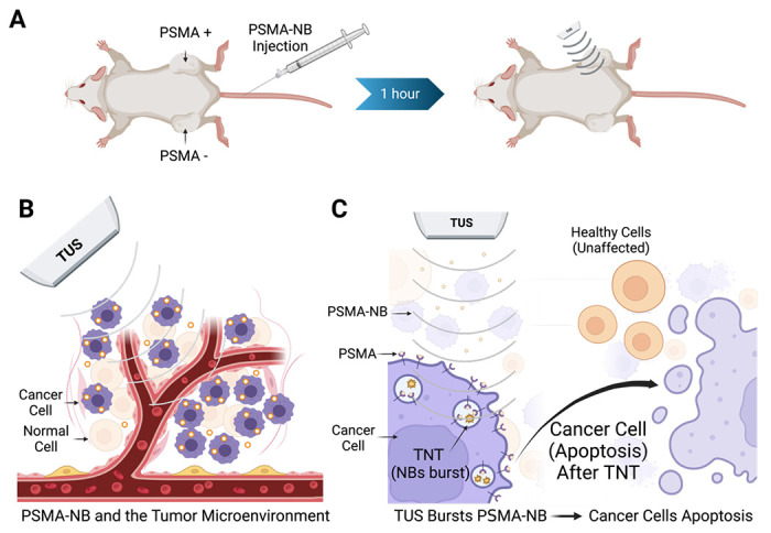
Lipid-shelled nanobubbles (NBs) can be visualized and activated using noninvasive ultrasound (US) stimulation, leading to significant bioeffects. We have previously shown that active targeting of NBs to prostate-specific membrane antigen (PSMA) overexpressed in prostate cancer (PCa) enhances the cellular internalization and prolongs retention of NBs with persistent acoustic activity (~hrs.). In this work, we hypothesized that tumor-accumulated PSMA-NBs combined with low frequency therapeutic US (TUS) will lead to selective damage and induce a therapeutic effect in PSMA-expressing tumors compared to PSMA-negative tumors.
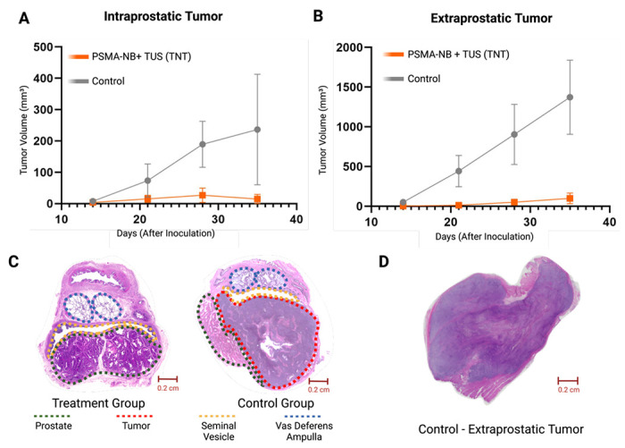
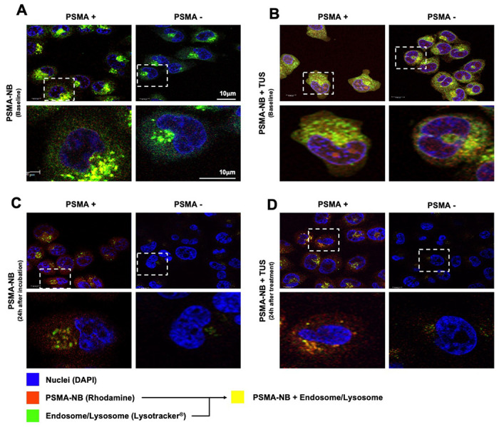
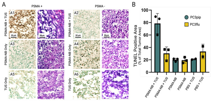
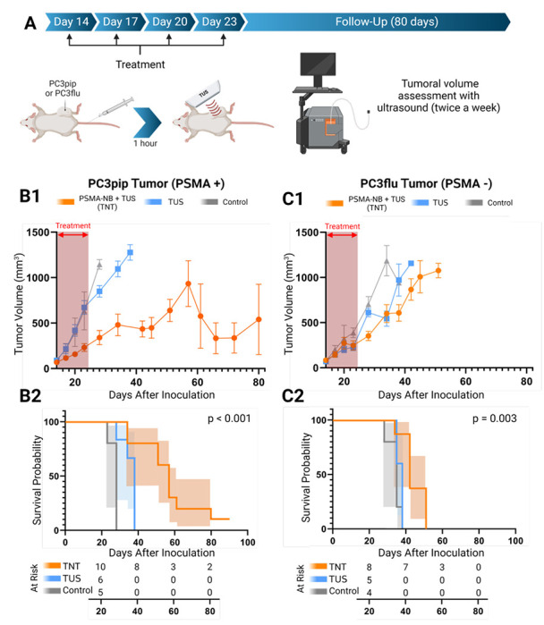
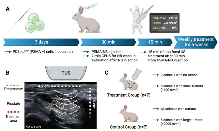
PSMA-targeted NBs were formulated by following our previously established protocol. Cellular internalization of fluorescent PSMA-NBs was evaluated by confocal imaging using late endosome/lysosome staining pre- and post-TUS application. Two animal models were used to assess the technique. Mice with dual tumors (PSMA expressing and PSMA negative) received PSMA-NB injection via the tail vein followed by TUS 1 hr. post injection (termed, targeted NB therapy or TNT). Twenty-four hours after treatment mice were euthanized and tumor cell apoptosis evaluated via TUNEL staining. Mice with single tumors (either PSMA + or -) were used for survival studies. Tumor size was measured for 80 days after four consecutive TNT treatments (every 3 days). To test the approach in a larger model, immunosuppressed rabbits with orthotopic human PSMA expressing tumors received PSMA-NB injection via the tail vein followed by TUS 30 min after injection. Tumor progression was assessed via US imaging and at the end point apoptosis was measured via TUNEL staining.
In vitro TNT studies using confocal microscopy showed that the internalized NBs and cellular compartments were disrupted after the TUS application, yet treated cells remained intact and viable. In vivo, PSMA-expressing tumors in mice receiving TNT treatment demonstrated a significantly greater extent of apoptosis (78.45 ± 9.3%, p < 0.01) compared to the other groups. TNT treatment significantly inhibited the PSMA (+) tumor growth and overall survival significantly improved (median survival time increase by 103%, p < 0.001). A significant reduction in tumor progression compared to untreated control was also seen in the rabbit model in intraprostatic (90%) and in extraprostatic lesions (94%) (p = 0.069 and 0.003, respectively).
We demonstrate for the first time the effect of PSMA-targeted nanobubble intracellular cavitation on cancer cell viability and tumor progression in two animal models. Data demonstrate that the targeted nanobubble therapy (TNT) approach relies primarily on mechanical disruption of intracellular vesicles and the resulting bioeffects appear to be more specific to target cancer cells expressing the PSMA receptor. The effect, while not lethal , resulted in significant tumor apoptosis in both a mouse and a rabbit model of PCa. While the mechanism of action of these effects is yet unclear, it is likely related to a locally-induced immune response, opening the door to future investigations in this area.
脂质包裹的纳米气泡(NBs)可通过无创超声(US)刺激进行可视化和激活,从而产生显著的生物效应。我们之前已经表明,将NBs主动靶向前列腺癌(PCa)中过表达的前列腺特异性膜抗原(PSMA),可增强细胞对NBs的内化作用,并延长具有持续声学活性(约数小时)的NBs的滞留时间。在这项研究中,我们假设与PSMA阴性肿瘤相比,肿瘤内积聚的PSMA-NBs联合低频治疗性超声(TUS)将导致PSMA表达肿瘤的选择性损伤并诱导治疗效果。
按照我们之前建立的方案制备PSMA靶向的NBs。使用共聚焦成像通过在TUS应用前后对晚期内体/溶酶体进行染色来评估荧光PSMA-NBs的细胞内化情况。使用两种动物模型来评估该技术。患有双肿瘤(PSMA表达和PSMA阴性)的小鼠通过尾静脉注射PSMA-NB,然后在注射后1小时进行TUS(称为靶向NB治疗或TNT)。治疗24小时后对小鼠实施安乐死,并通过TUNEL染色评估肿瘤细胞凋亡情况。患有单肿瘤(PSMA阳性或阴性)的小鼠用于生存研究。在连续进行四次TNT治疗(每3天一次)后的80天内测量肿瘤大小。为了在更大的模型中测试该方法,对患有原位人PSMA表达肿瘤的免疫抑制兔通过尾静脉注射PSMA-NB,然后在注射后30分钟进行TUS。通过超声成像评估肿瘤进展情况,并在终点时通过TUNEL染色测量凋亡情况。
使用共聚焦显微镜进行的体外TNT研究表明,在TUS应用后,内化的NBs和细胞区室被破坏,但处理后的细胞仍保持完整且存活。在体内,接受TNT治疗的小鼠中表达PSMA的肿瘤与其他组相比,凋亡程度显著更高(78.45±9.3%,p<0.01)。TNT治疗显著抑制了PSMA(+)肿瘤的生长,总体生存率显著提高(中位生存时间增加103%,p<0.001)。在兔模型中,与未治疗的对照组相比,前列腺内(90%)和前列腺外病变(94%)的肿瘤进展也显著降低(分别为p = 0.069和0.003)。
我们首次在两种动物模型中证明了PSMA靶向纳米气泡细胞内空化对癌细胞活力和肿瘤进展的影响。数据表明,靶向纳米气泡治疗(TNT)方法主要依赖于细胞内囊泡的机械破坏,并且所产生的生物效应似乎对表达PSMA受体的靶癌细胞更具特异性。这种效应虽然不致命,但在PCa的小鼠和兔模型中均导致了显著的肿瘤凋亡。虽然这些效应的作用机制尚不清楚,但可能与局部诱导的免疫反应有关,为该领域的未来研究打开了大门。