Sauter Andreas P, Proksa Roland, Knipfer Andreas, Reischl Stefan, Braren Rickmer F, Nadjiri Jonathan, Kopp Felix, Noël Peter B, Makowski Markus R, Rummeny Ernst J, Fingerle Alexander A
Department of Diagnostic and Interventional Radiology, School of Medicine and Klinikum Rechts Der Isar, Technical University of Munich, Ismaningerstr. 22, 81675, Munich, Germany.
Philips Research, Hamburg, Germany.
Cardiovasc Intervent Radiol. 2023 Nov;46(11):1621-1631. doi: 10.1007/s00270-023-03550-7. Epub 2023 Sep 27.
Evaluation of dual-layer spectral computed tomography (CT) for contrast enhancement during image-guided biopsy of liver lesions using virtual monoenergetic images (VMI) and virtual non-contrast (VNC) images.
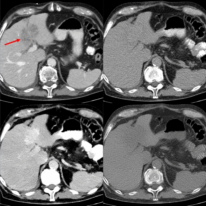
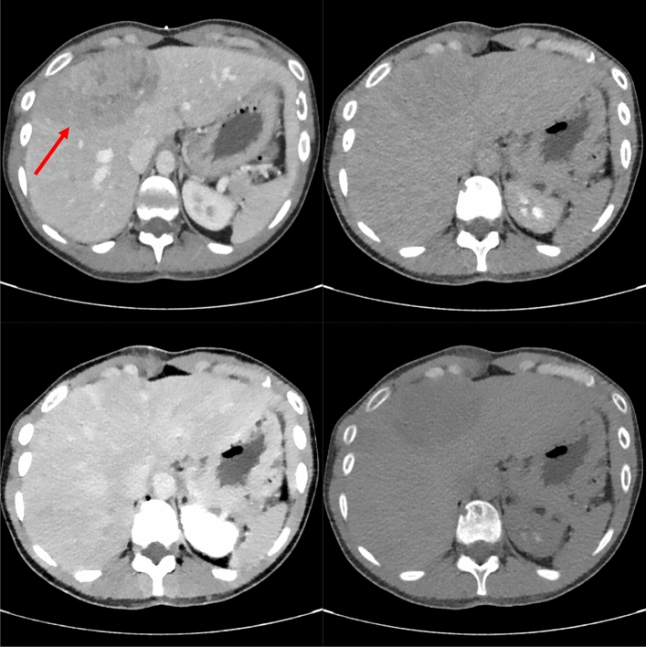
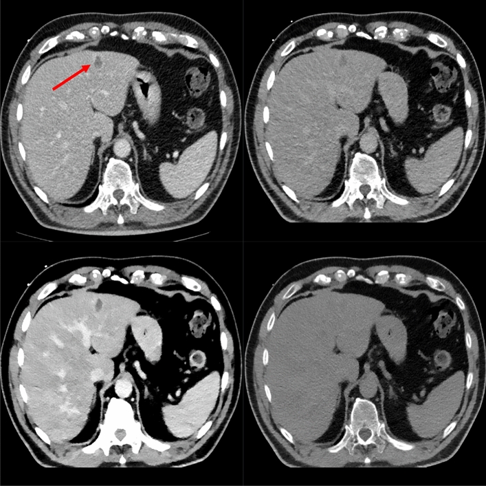
Spectral CT data of 20 patients receiving CT-guided needle biopsy of focal liver lesions were used to generate VMI at energy levels from 40 to 200 keV and VNC images. Images were analyzed objectively regarding contrast-to-noise ratio between lesion center (CNR) or periphery (CNR) and normal liver parenchyma. Lesion visibility and image quality were evaluated on a 4-point Likert scale by two radiologists.
Using VMI/VNC images, readers reported an increased visibility of the lesion compared to the conventional CT images in 18/20 cases. In 75% of cases, the highest visibility was derived by VMI-40. Showing all reconstructions simultaneously, VMI-40 offered the highest visibility in 75% of cases, followed by VNC in 12.5% of cases. Either CNR (17/20) or/and CNR (17/20) was higher (CNR increase > 50%) in 19/20 cases for VMI-40 or VNC images compared to conventional CT images. VMI-40 showed the highest CNR in 14 cases and the highest CNR in 12 cases. High image quality was present for all reconstructions with a minimum median of 3.5 for VMI-40 and VMI-50.
When implemented in the CT scanner software, automated contrast enhancement of liver lesions during image-guided biopsy may facilitate the procedure.
使用虚拟单能量图像(VMI)和虚拟平扫(VNC)图像评估双层光谱计算机断层扫描(CT)在肝脏病变图像引导活检过程中的对比增强效果。
对20例接受CT引导下局灶性肝脏病变穿刺活检的患者的光谱CT数据进行分析,以生成40至200keV能量水平的VMI和VNC图像。客观分析病变中心(CNR)或周边(CNR)与正常肝实质之间的对比噪声比。由两名放射科医生采用4分李克特量表对病变可视性和图像质量进行评估。
使用VMI/VNC图像时,与传统CT图像相比,在18/20例病例中,阅片者报告病变的可视性有所提高。在75%的病例中,VMI-40的可视性最高。同时显示所有重建图像时,VMI-40在75%的病例中可视性最高,其次是VNC,占12.5%的病例。与传统CT图像相比,在19/20例病例中,VMI-40或VNC图像的CNR(17/20)或/和CNR(17/20)更高(CNR增加>50%)。VMI-40在14例中显示出最高的CNR,在12例中显示出最高的CNR。所有重建图像的图像质量都很高,VMI-40和VMI-50的最低中位数为3.5。
当在CT扫描仪软件中实施时,图像引导活检过程中肝脏病变的自动对比增强可能会促进该操作。