Wang Xinmei, Shuai Guangnan, Xu Junhui, Liu Meihua, Zhao Jianguo, Zhang Na, Zhang Wenwen, Qu Pengpeng
Department of Gynecological Oncology, Tianjin Central Hospital of Gynecology and Obstetrics, Tianjin, China.
Wenjiang District People's Hospital of Chengdu, Chengdu, Sichuan, China.
Front Oncol. 2023 Sep 15;13:1221962. doi: 10.3389/fonc.2023.1221962. eCollection 2023.
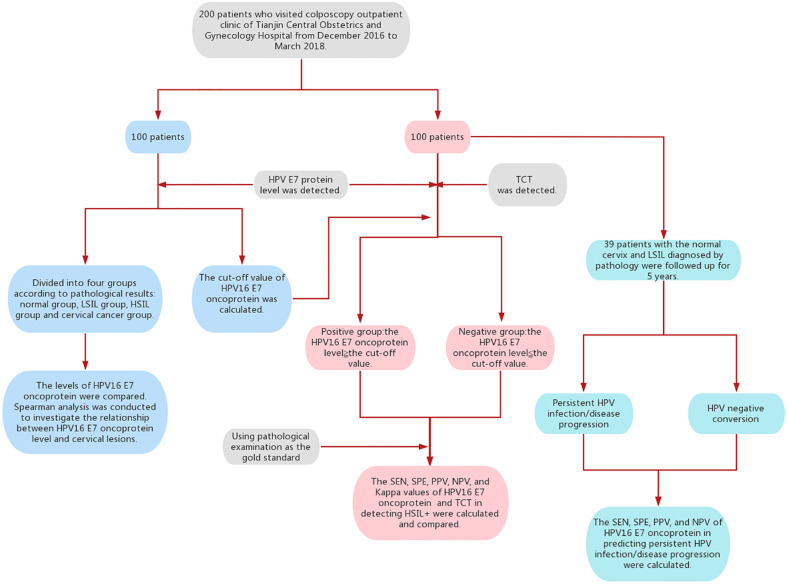
Colposcopy is recommended once human papillomavirus (HPV)16/18 infection is detected. However, not all HPV16/18-positive women will necessarily develop cervical lesions. Therefore, this study aimed to investigate the application of quantitative HPV16 E7 oncoprotein detection as a cervical cancer screening method for more efficient screening while minimizing unnecessary colposcopy.
E7 oncoprotein (HPV16) was quantitatively detected in cervical exfoliated cells of HPV16-positive women. The levels of HPV16 E7 oncoprotein in different degrees of cervical lesions were compared, and the optimal cut-off value for identifying HSIL+ was determined by receiver operating characteristic (ROC) curve analysis. With a pathological diagnosis as the gold standard, the sensitivity (SEN), specificity (SPE), positive predictive value (PPV), negative predictive value (NPV), and Kappa value were calculated to verify the diagnostic value of the method. Women diagnosed with low-grade squamous intraepithelial lesions (LSIL) and normal women were followed up for 5 years to evaluate the predictive value of HPV16 E7 protein for disease progression/persistent infection.
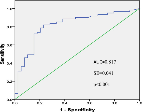
The expression level of HPV16 E7 oncoprotein was positively correlated with the degree of the cervical lesion (r = 0.589, P < 0.01). The area under the ROC curve (AUC) was 0.817 (confidence interval: 0.729-0.904). The cut-off value of E7 oncoprotein for identifying HSIL+ was 8.68 ng/ml. The SEN, SPE, PPV, NPV, and Kappa values of HPV16 E7 oncoprotein for the identification of HSIL+ were 87.1%,70.0%, 87.1%, 70.0%, and 0.571, respectively, which were higher than those of ThinPrep cytology test (TCT). The SEN, SPE, PPV, and NPV of HPV16 E7 oncoprotein in predicting disease progression/persistent infection were 93.75%, 91.30%, 88.24%, and 95.45%, respectively.
The quantitative detection of HPV 16 E7 oncoprotein can not only accurately screen cervical lesions but also achieve efficient colposcopy referral. Additionally, HPV16 E7 oncoprotein can accurately predict the progression of cervical lesions and persistent HPV infection.
一旦检测到人类乳头瘤病毒(HPV)16/18感染,建议进行阴道镜检查。然而,并非所有HPV16/18阳性女性都会发展为宫颈病变。因此,本研究旨在探讨定量检测HPV16 E7癌蛋白作为一种宫颈癌筛查方法的应用,以实现更有效的筛查,同时尽量减少不必要的阴道镜检查。
对HPV16阳性女性的宫颈脱落细胞进行HPV16 E7癌蛋白定量检测。比较不同程度宫颈病变中HPV16 E7癌蛋白水平,通过受试者工作特征(ROC)曲线分析确定识别高级别鳞状上皮内病变(HSIL+)的最佳临界值。以病理诊断为金标准,计算灵敏度(SEN)、特异度(SPE)、阳性预测值(PPV)、阴性预测值(NPV)和Kappa值,以验证该方法的诊断价值。对诊断为低级别鳞状上皮内病变(LSIL)的女性和正常女性进行5年随访,以评估HPV16 E7蛋白对疾病进展/持续感染的预测价值。
HPV16 E7癌蛋白表达水平与宫颈病变程度呈正相关(r = 0.589,P < 0.01)。ROC曲线下面积(AUC)为0.817(置信区间:0.729 - 0.904)。识别HSIL+的E7癌蛋白临界值为8.68 ng/ml。HPV16 E7癌蛋白识别HSIL+的SEN、SPE、PPV、NPV和Kappa值分别为87.1%、70.0%、87.1%、70.0%和0.571,高于薄层液基细胞学检测(TCT)。HPV16 E7癌蛋白预测疾病进展/持续感染的SEN、SPE、PPV和NPV分别为93.75%、91.30%、88.24%和95.45%。
HPV 16 E7癌蛋白定量检测不仅能准确筛查宫颈病变,还能实现有效的阴道镜转诊。此外,HPV16 E7癌蛋白能准确预测宫颈病变进展和HPV持续感染。