Department of Stomatology, General Hospital of Ningxia Medical University, Yinchuan, China.
Institute of Stem Cells, General Hospital of Ningxia Medical University, Yinchuan, China.
Cell Transplant. 2023 Jan-Dec;32:9636897231202541. doi: 10.1177/09636897231202541.
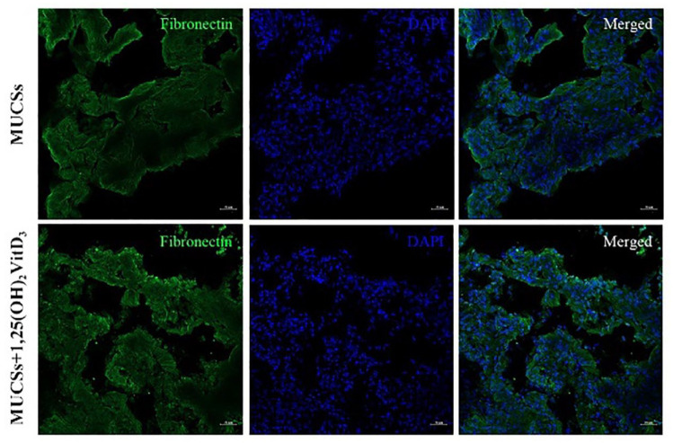
This study aims to investigate the effects of 1,25-dihydroxyvitamin D3 (1,25(OH)VitD) on osteogenic differentiation of human periodontal ligament stem cells (hPDLSCs) and the activity of hPDLSC sheets and the differences in the tissue regeneration activity of hPDLSC sheets on tooth root fragment treated by different methods. Healthy caries-free premolars were collected. The hPDLSCs were obtained by enzymatic digestion. Surface markers of stem cells were analyzed by flow cytometry and the multidirectional differentiation ability of hPDLSCs was detected. During the osteogenic differentiation of hPDLSCs, 1,25(OH)VitD was added and the effect of 1,25(OH)VitD on osteogenic differentiation of hPDLSCs was assessed using Western blotting, quantitative reverse transcription-polymerase chain reaction (qRT-PCR), enzyme-linked immunosorbent assay, cell staining, and immunofluorescence. After hPDLSC sheets were prepared, histology and immunofluorescence analysis of the effect of 1,25(OH)VitD on sheet activity were performed. In addition, root fragments were prepared and treated with scaling, 24% EDTA (ethylenediamide tetraacetic acid), and Er,Cr:YSGG lasers, respectively, and the tissue regeneration activity of hPDLSC sheets on different root fragments were observed. 1,25(OH)VitD promoted the high gene and protein expressions of osteogenic markers ALP (alkaline phosphatase), Runx2, and OPN (osteopontin antibody) in hPDLSCs, along with enhanced ALP activity and staining, alizarin red staining, and immunofluorescence staining, indicating that the osteogenic differentiation ability of hPDLSCs was improved. Extracellular matrix secretion was increased in hPDLSC sheets, along with the positive expressions of the protein markers fibronectin and collagen I, suggesting that 1,25(OH)VitD could enhance these effects. In addition, the root fragments treated by Er,Cr:YSGG laser were more suitable for the attachment and regeneration of hPDLSC sheets, demonstrating that 1,25(OH)VitD could improve the tissue regeneration performance of these sheets. 1,25(OH)VitD can promote osteogenic differentiation of hPDLSCs and thus plays an active role in hPDLSC sheet formation and tissue regeneration. In addition, the Er,Cr:YSGG laser can be used as the recommended treatment method for the root surface regenerated by hPDLSCs.
本研究旨在探讨 1,25-二羟维生素 D3(1,25(OH)VitD)对人牙周膜干细胞(hPDLSCs)成骨分化的影响,以及 hPDLSC 片的活性,以及不同方法处理的牙根碎片上 hPDLSC 片的组织再生活性的差异。收集健康无龋前磨牙。通过酶消化获得 hPDLSCs。通过流式细胞术分析干细胞表面标志物,并检测 hPDLSCs 的多向分化能力。在 hPDLSCs 成骨分化过程中,加入 1,25(OH)VitD,通过 Western blot、定量逆转录-聚合酶链反应(qRT-PCR)、酶联免疫吸附试验、细胞染色和免疫荧光评估 1,25(OH)VitD 对 hPDLSCs 成骨分化的影响。制备 hPDLSC 片后,进行组织学和免疫荧光分析 1,25(OH)VitD 对片活性的影响。此外,分别对牙根碎片进行洁治、24% EDTA(乙二胺四乙酸)和 Er、Cr:YSGG 激光处理,观察 hPDLSC 片在不同牙根碎片上的组织再生活性。1,25(OH)VitD 促进 hPDLSCs 中碱性磷酸酶(ALP)、Runx2 和骨桥蛋白抗体(OPN)等成骨标志物的高基因和蛋白表达,并增强 ALP 活性和染色、茜素红染色和免疫荧光染色,表明 hPDLSCs 的成骨分化能力得到提高。hPDLSC 片的细胞外基质分泌增加,同时纤维连接蛋白和 I 型胶原等蛋白标志物的表达呈阳性,表明 1,25(OH)VitD 可以增强这些作用。此外,经 Er、Cr:YSGG 激光处理的牙根碎片更适合 hPDLSC 片的附着和再生,表明 1,25(OH)VitD 可以提高这些片的组织再生性能。1,25(OH)VitD 可促进 hPDLSCs 的成骨分化,从而在 hPDLSC 片的形成和组织再生中发挥积极作用。此外,Er、Cr:YSGG 激光可用作 hPDLSCs 再生根表面的推荐治疗方法。