Jaha Luan, Ademi Bekim, Rudari Hajriz, Vokrri Lulzim, Gjikolli Bujar, Koshi Adhurim, Kuçi Astrit, Jaha Art
Department of Vascular Surgery University Clinical Center of Kosovo Prishtina Kosovo.
Department of Radiology University Clinical Center of Kosovo Prishtina Kosovo.
Clin Case Rep. 2023 Oct 3;11(10):e8015. doi: 10.1002/ccr3.8015. eCollection 2023 Oct.
Extracranial internal carotid artery aneurysms (EICAAs) can lead to serious medical conditions, such as stroke or compression over cranial nerves. In very few cases, there may be hemorrhagic complications due to the rupture. Although rare, they should be suspected cause in every patient with transitory ischemic attack or stroke, especially in the presence of pain, palpable mass or bruit in the neck.
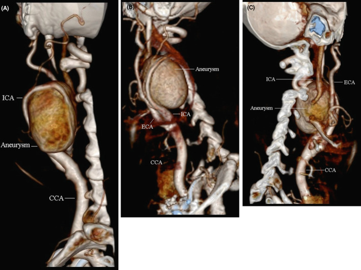
Extracranial internal carotid artery aneurysms (EICAAs) are rare arterial aneurysms, with a prevalence of less than 1%. Although they are not common, these aneurysms can lead to serious medical conditions, such as stroke or compression over cranial nerves. In very few cases, there may be hemorrhagic complications due to the rupture of the aneurysm. This report presents a case of a successful surgical intervention for EICAA, and an overview of symptoms, risk factors, causes, diagnostic procedures, treatments, and potential postoperative complications. A 70-year-old Albanian lady had been experiencing pain due to a pulsating mass in her neck for many years. Physical examination did not reveal any signs of infection, injury, or previous surgery. A palpable thrill and a carotid bruit were detected over an evident pulsating mass on the left side of her neck. Her past medical history was consistent with three transitory ischemic attacks in recent months and a stroke 5 years earlier. Comorbidities included hypercholesterolemia, hypertension, and long-standing coronary artery disease. Imaging investigation in terms of ultrasound and CT-scan confirmed the presence of an aneurysm of the proximal tract of the internal carotid artery measuring 42 × 31 mm. Surgery was indicated on symptomatic and anatomical grounds. The procedure was carried out under general anesthesia. After proximal and distal clamping, the aneurysm was excised followed by end-to-end anastomosis of the internal carotid artery. The postoperative course was uneventful, and the patient was discharged home on the fifth postoperative day. Despite the growing number of reported cases of successful endovascular treatment for internal carotid artery aneurysms, open surgery remains a safe and effective treatment option. However, it is crucial to provide customized treatment plans for each patient based on their individual characteristics and the particularities of their aneurysm.
颅外颈内动脉瘤(EICAA)可导致严重的医疗状况,如中风或压迫颅神经。在极少数情况下,动脉瘤破裂可能会引发出血并发症。虽然罕见,但对于每一位短暂性脑缺血发作或中风患者,尤其是伴有颈部疼痛、可触及肿块或血管杂音时,都应怀疑存在EICAA。
颅外颈内动脉瘤(EICAA)是罕见的动脉性动脉瘤,患病率低于1%。尽管并不常见,但这些动脉瘤可导致严重的医疗状况,如中风或压迫颅神经。在极少数情况下,动脉瘤破裂可能会引发出血并发症。本报告介绍了一例成功进行手术干预治疗EICAA的病例,并概述了其症状、危险因素、病因、诊断程序、治疗方法以及潜在的术后并发症。一名70岁的阿尔巴尼亚女性多年来一直因颈部搏动性肿块而感到疼痛。体格检查未发现任何感染、损伤或既往手术的迹象。在其颈部左侧明显的搏动性肿块上检测到可触及的震颤和颈动脉杂音。她的既往病史显示,近几个月有三次短暂性脑缺血发作,5年前曾发生过一次中风。合并症包括高胆固醇血症、高血压和长期的冠状动脉疾病。超声和CT扫描等影像学检查证实颈内动脉近端存在一个大小为42×31mm的动脉瘤。基于症状和解剖学原因,建议进行手术。手术在全身麻醉下进行。在近端和远端夹闭后,切除动脉瘤,随后进行颈内动脉端端吻合。术后过程顺利,患者于术后第五天出院。尽管报道的成功进行颈内动脉瘤血管内治疗的病例数量不断增加,但开放手术仍然是一种安全有效的治疗选择。然而,根据每位患者的个体特征和动脉瘤的特殊性提供定制化的治疗方案至关重要。