• 文献检索
  • 文档翻译
  • 深度研究
  • 学术资讯
  • Suppr Zotero 插件Zotero 插件
  • 邀请有礼
  • 套餐&价格
  • 历史记录
应用&插件
Suppr Zotero 插件Zotero 插件浏览器插件Mac 客户端Windows 客户端微信小程序
定价
高级版会员购买积分包购买API积分包
服务
文献检索文档翻译深度研究API 文档MCP 服务
关于我们
关于 Suppr公司介绍联系我们用户协议隐私条款
关注我们

Suppr 超能文献

核心技术专利:CN118964589B侵权必究
粤ICP备2023148730 号-1Suppr @ 2026

文献检索

告别复杂PubMed语法,用中文像聊天一样搜索,搜遍4000万医学文献。AI智能推荐,让科研检索更轻松。

立即免费搜索

文件翻译

保留排版,准确专业,支持PDF/Word/PPT等文件格式,支持 12+语言互译。

免费翻译文档

深度研究

AI帮你快速写综述,25分钟生成高质量综述,智能提取关键信息,辅助科研写作。

立即免费体验

载脂蛋白 B 与 LDL-C 比值在动脉粥样硬化斑块中的作用及其与心血管事件的关系

Association of LDL-C level with neoatherosclerosis and plaque vulnerability in patients with late restenosis: an optical coherence tomography study.

机构信息

Department of Cardiology, Affiliated Hospital of Zunyi Medical University, Zunyi, 563000, China.

Department of Cardiology, the Fifth Affiliated Hospital of Zunyi Medical University, Zhuhai, China.

出版信息

Int J Cardiovasc Imaging. 2023 Dec;39(12):2609-2619. doi: 10.1007/s10554-023-02956-1. Epub 2023 Oct 7.

DOI:10.1007/s10554-023-02956-1
PMID:37804387
原文链接:https://pmc.ncbi.nlm.nih.gov/articles/PMC10691981/
Abstract

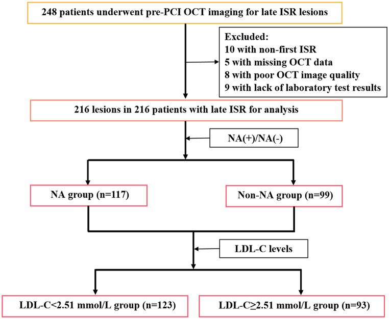
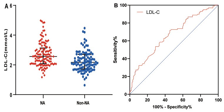
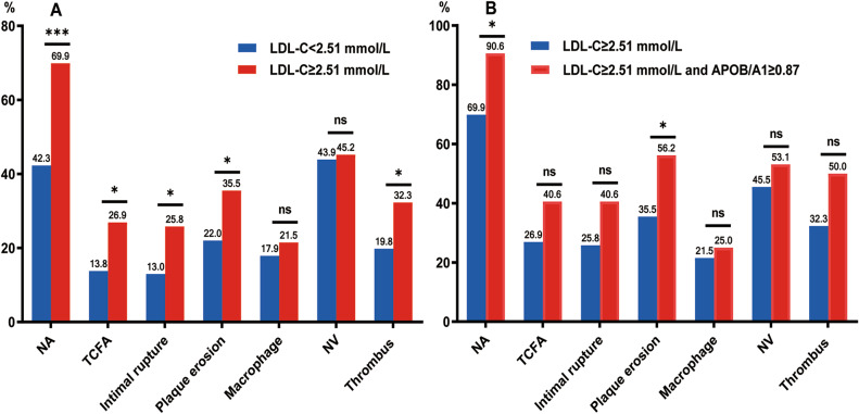
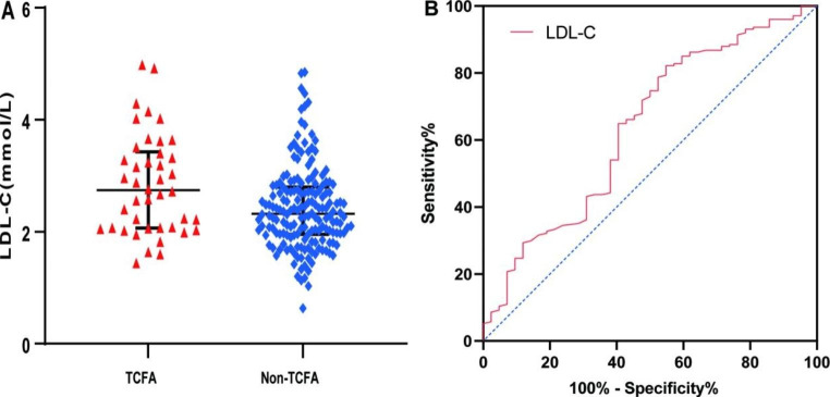
Neoatherosclerosis (NA) is a significant contributor to late stent failure; however, predictors of late in-stent restenosis (ISR) with NA have not been systematically reported. This study aimed to identify predictors of NA incidence and plaque vulnerability in patients with late ISR and the role of low-density lipoprotein cholesterol (LDL-C) levels in this process. A total of 216 patients with 216 lesions who underwent optical coherence tomography (OCT) before interventional procedure for late drug-eluting stent ISR were enrolled and divided into NA and non-NA groups based on OCT findings. Results showed that higher LDL-C levels were associated with NA, thin-cap fibroatheroma (TCFA), intimal disruption, plaque erosion, and thrombosis. Multivariate regression analysis revealed that the LDL-C level was an independent risk factor for NA and TCFA. The LDL-C levels exhibited a significant predictive value for NA and TCFA, surpassing other factors such as stent age and other lipid types. In conclusion, a high LDL-C level is an independent predictor of NA incidence and plaque vulnerability in patients with late ISR.

摘要

动脉新生内膜形成(NA)是导致晚期支架失败的重要原因,但目前尚未系统报道与 NA 相关的晚期支架内再狭窄(ISR)的预测因素。本研究旨在确定晚期 ISR 患者发生 NA 和易损斑块的预测因素,以及低密度脂蛋白胆固醇(LDL-C)水平在这一过程中的作用。共纳入 216 例因晚期药物洗脱支架 ISR 而行介入治疗前接受光学相干断层扫描(OCT)检查的患者,根据 OCT 结果分为 NA 组和非 NA 组。结果显示,较高的 LDL-C 水平与 NA、薄帽纤维粥样斑块(TCFA)、内膜撕裂、斑块侵蚀和血栓形成有关。多变量回归分析显示,LDL-C 水平是 NA 和 TCFA 的独立危险因素。LDL-C 水平对 NA 和 TCFA 具有显著的预测价值,超过了支架年龄和其他脂质类型等其他因素。总之,高 LDL-C 水平是晚期 ISR 患者发生 NA 和易损斑块的独立预测因素。

https://cdn.ncbi.nlm.nih.gov/pmc/blobs/a08e/10691981/5716ca40aff9/10554_2023_2956_Fig5_HTML.jpg
https://cdn.ncbi.nlm.nih.gov/pmc/blobs/a08e/10691981/f37c7257967d/10554_2023_2956_Fig1_HTML.jpg
https://cdn.ncbi.nlm.nih.gov/pmc/blobs/a08e/10691981/6f54ba72a16c/10554_2023_2956_Fig2_HTML.jpg
https://cdn.ncbi.nlm.nih.gov/pmc/blobs/a08e/10691981/8d12c0e1e425/10554_2023_2956_Fig3_HTML.jpg
https://cdn.ncbi.nlm.nih.gov/pmc/blobs/a08e/10691981/6d9fe5a22fbd/10554_2023_2956_Fig4_HTML.jpg
https://cdn.ncbi.nlm.nih.gov/pmc/blobs/a08e/10691981/5716ca40aff9/10554_2023_2956_Fig5_HTML.jpg

相似文献

1
Association of LDL-C level with neoatherosclerosis and plaque vulnerability in patients with late restenosis: an optical coherence tomography study.载脂蛋白 B 与 LDL-C 比值在动脉粥样硬化斑块中的作用及其与心血管事件的关系
Int J Cardiovasc Imaging. 2023 Dec;39(12):2609-2619. doi: 10.1007/s10554-023-02956-1. Epub 2023 Oct 7.
2
Optical coherence tomographic analysis of drug-eluting in-stent restenosis at different times: A STROBE compliant study.不同时间药物洗脱支架内再狭窄的光学相干断层扫描分析:一项遵循STROBE规范的研究
Medicine (Baltimore). 2018 Aug;97(34):e12117. doi: 10.1097/MD.0000000000012117.
3
Association of the monocytes to high-density lipoprotein cholesterol ratio with in-stent neoatherosclerosis and plaque vulnerability: An optical coherence tomography study.单核细胞/高密度脂蛋白胆固醇比值与支架内新动脉粥样硬化及斑块易损性的相关性:一项光学相干断层成像研究。
Int J Cardiol. 2024 Feb 1;396:131417. doi: 10.1016/j.ijcard.2023.131417. Epub 2023 Oct 4.
4
Comparison of in-stent neoatherosclerosis and tissue characteristics between early and late in-stent restenosis in second-generation drug-eluting stents: an optical coherence tomography study.第二代药物洗脱支架内早期与晚期支架内再狭窄的支架内新生动脉粥样硬化及组织特征比较:一项光学相干断层扫描研究
Int J Cardiovasc Imaging. 2017 Oct;33(10):1463-1472. doi: 10.1007/s10554-017-1146-7. Epub 2017 Apr 25.
5
Lipoprotein (a) is related to In-Stent neoatherosclerosis incidence rate and plaque vulnerability: Optical Coherence Tomography Study.脂蛋白(a)与支架内新动脉粥样硬化发生率及斑块易损性相关:光学相干断层成像研究。
Int J Cardiovasc Imaging. 2023 Feb;39(2):275-284. doi: 10.1007/s10554-022-02736-3. Epub 2022 Oct 31.
6
Optical coherence tomographic analysis of in-stent neoatherosclerosis after drug-eluting stent implantation.药物洗脱支架置入后支架内新动脉粥样硬化的光学相干断层成像分析。
Circulation. 2011 Jun 28;123(25):2954-63. doi: 10.1161/CIRCULATIONAHA.110.988436. Epub 2011 Jun 6.
7
Naturally Occurring Atherosclerosis Progression and In-stent Restenosis: Exploring Histomorphologic Associations Using Optical Coherence Tomography.自然发生的动脉粥样硬化进展和支架内再狭窄:应用光学相干断层成像技术探索组织形态学相关性。
J Cardiovasc Pharmacol. 2024 Jun 1;83(6):646-654. doi: 10.1097/FJC.0000000000001520.
8
[Optical coherence tomographic analysis of in-stent neoatherosclerosis in lesions with restenosis after drug-eluting stent implantation].[药物洗脱支架植入术后再狭窄病变中支架内新生动脉粥样硬化的光学相干断层扫描分析]
Zhonghua Xin Xue Guan Bing Za Zhi. 2018 Jan 24;46(1):44-49. doi: 10.3760/cma.j.issn.0253-3758.2018.01.008.
9
Potential Mechanisms of In-stent Neointimal Atherosclerotic Plaque Formation.支架内新生动脉粥样硬化斑块形成的潜在机制。
J Cardiovasc Pharmacol. 2021 Sep 1;78(3):388-393. doi: 10.1097/FJC.0000000000001059.
10
Impact of baseline plaque characteristic on the development of neoatherosclerosis in the very late phase after stenting.支架置入后极晚期新生动脉粥样硬化中基线斑块特征的影响。
J Cardiol. 2019 Jul;74(1):67-73. doi: 10.1016/j.jjcc.2019.01.002. Epub 2019 Feb 4.

引用本文的文献

1
Predictive Utility of PLR and Platelet-to-LDL Ratio for in-Stent Restenosis Following Carotid Artery Stenting.PLR和血小板与低密度脂蛋白比值对颈动脉支架置入术后支架内再狭窄的预测效用
Vasc Health Risk Manag. 2025 Aug 27;21:671-684. doi: 10.2147/VHRM.S534400. eCollection 2025.
2
Neovascularization restructuring patterns in diabetic patients with coronary in stent restenosis: an in-vivo optical coherence tomography study.糖尿病合并冠状动脉支架内再狭窄患者的新生血管重构模式:一项体内光学相干断层扫描研究
Int J Cardiovasc Imaging. 2025 Feb;41(2):225-235. doi: 10.1007/s10554-024-03301-w. Epub 2024 Dec 19.
3
Impact of acute coronary syndrome on early in-stent neoatherosclerosis as shown by optical coherence tomography.

本文引用的文献

1
Impact of triglyceride-rich lipoproteins on early in-stent neoatherosclerosis formation in patients undergoing statin treatment.富含甘油三酯的脂蛋白对接受他汀类药物治疗患者早期支架内新生动脉粥样硬化形成的影响。
J Clin Lipidol. 2023 Mar-Apr;17(2):281-290. doi: 10.1016/j.jacl.2023.01.004. Epub 2023 Jan 26.
2
Global research trends in in-stent neoatherosclerosis: A CiteSpace-based visual analysis.支架内新生动脉粥样硬化的全球研究趋势:基于CiteSpace的可视化分析
Front Cardiovasc Med. 2022 Nov 8;9:1025858. doi: 10.3389/fcvm.2022.1025858. eCollection 2022.
3
Lipoprotein (a) is related to In-Stent neoatherosclerosis incidence rate and plaque vulnerability: Optical Coherence Tomography Study.
光学相干断层扫描显示急性冠状动脉综合征对早期支架内新生动脉粥样硬化的影响。
AsiaIntervention. 2024 Sep 27;10(3):203-211. doi: 10.4244/AIJ-D-24-00021. eCollection 2024 Sep.
4
Neoatherosclerosis: A Distinctive Pathological Mechanism of Stent Failure.新型动脉粥样硬化:支架失败的独特病理机制
Rev Cardiovasc Med. 2024 Mar 7;25(3):95. doi: 10.31083/j.rcm2503095. eCollection 2024 Mar.
脂蛋白(a)与支架内新动脉粥样硬化发生率及斑块易损性相关:光学相干断层成像研究。
Int J Cardiovasc Imaging. 2023 Feb;39(2):275-284. doi: 10.1007/s10554-022-02736-3. Epub 2022 Oct 31.
4
Prevalence and Impact of Neoatherosclerosis on Clinical Outcomes After Percutaneous Treatment of Second-Generation Drug-Eluting Stent Restenosis.第二代药物洗脱支架再狭窄经皮治疗后新生动脉粥样硬化的流行及对临床结局的影响。
Circ Cardiovasc Interv. 2022 Sep;15(9):e011693. doi: 10.1161/CIRCINTERVENTIONS.121.011693. Epub 2022 Sep 20.
5
Optical coherence tomography in coronary atherosclerosis assessment and intervention.光学相干断层成像在冠状动脉粥样硬化评估和干预中的应用。
Nat Rev Cardiol. 2022 Oct;19(10):684-703. doi: 10.1038/s41569-022-00687-9. Epub 2022 Apr 21.
6
Association between apolipoprotein B/A1 ratio and coronary plaque vulnerability in patients with atherosclerotic cardiovascular disease: an intravascular optical coherence tomography study.载脂蛋白 B/A1 比值与动脉粥样硬化性心血管病患者冠状动脉斑块易损性的关系:一项血管内光学相干断层成像研究。
Cardiovasc Diabetol. 2021 Sep 15;20(1):188. doi: 10.1186/s12933-021-01381-9.
7
Thin-cap fibroatheroma predicts clinical events in diabetic patients with normal fractional flow reserve: the COMBINE OCT-FFR trial.薄帽纤维粥样斑块可预测正常血流储备分数的糖尿病患者的临床事件:COMBINE OCT-FFR 试验。
Eur Heart J. 2021 Dec 1;42(45):4671-4679. doi: 10.1093/eurheartj/ehab433.
8
Predictors and outcomes of neoatherosclerosis in patients with in-stent restenosis.支架内再狭窄患者的新生动脉粥样硬化的预测因素和结果。
EuroIntervention. 2021 Aug 27;17(6):489-496. doi: 10.4244/EIJ-D-20-00539.
9
Incidence and Predictors of Neoatherosclerosis in Patients with Early In-Stent Restenosis Determined Using Optical Coherence Tomography.使用光学相干断层扫描技术测定早期支架内再狭窄患者新动脉粥样硬化的发生率及预测因素。
Int Heart J. 2020 Sep 29;61(5):872-878. doi: 10.1536/ihj.20-139. Epub 2020 Sep 12.
10
Relationship between coronary plaque morphology of the left anterior descending artery and 12 months clinical outcome: the CLIMA study.左前降支冠状动脉斑块形态与 12 个月临床结局的关系:CLIMA 研究。
Eur Heart J. 2020 Jan 14;41(3):383-391. doi: 10.1093/eurheartj/ehz520.