Department of Radiology, Faculty of Medicine Siriraj Hospital, Mahidol University, 2 Wanglang Rd, Bangkok Noi, Bangkok, 10700, Thailand.
Department of Research, Faculty of Medicine Siriraj Hospital, Mahidol University, 2 Wanglang Rd, Bangkok Noi, Bangkok, 10700, Thailand.
Eur Radiol. 2024 Apr;34(4):2534-2545. doi: 10.1007/s00330-023-10273-3. Epub 2023 Oct 14.
Accurate computed tomography (CT) identification of appendicoliths in adults with acute appendicitis is crucial as it may preclude nonoperative management due to high risk of failure and complications. This investigation aimed to identify the significance of appendicoliths in acute appendicitis and to evaluate the performance of portovenous-phase (PVP) CT and the consequences of overlooked appendicoliths.
CT examinations of 324 consecutive patients (mean age 51.9 years, 112 men) with pathologically confirmed acute appendicitis were retrospectively included. Two radiologists independently reviewed the images, and disagreement was resolved by a consensus.
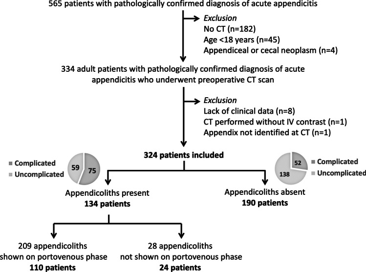
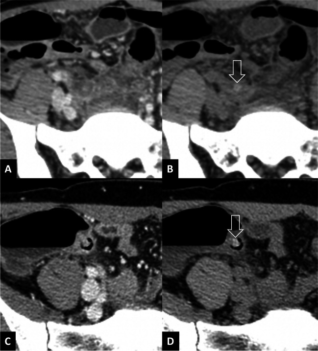
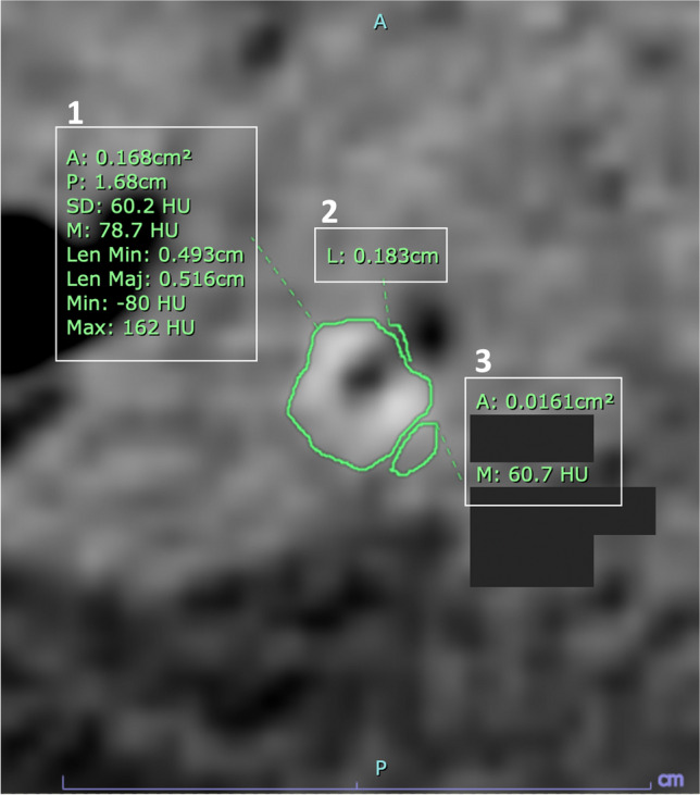
Appendicoliths were identified in 134/324 patients, of which 75 had complicated appendicitis. Among 190 patients without appendicoliths, 52 had complicated appendicitis. An appendicolith was independently associated with complicated appendicitis (adjusted odds ratio 2.289; 95% CI: 1.343-3.902; p = 0.002). The larger minimum diameter was significantly associated with complication. The 4.5-/6.0-mm cutoffs for minimum and maximum diameters of appendicoliths demonstrated 82.7%/85.3% sensitivity and 35.6%/33.9% specificity in predicting complications. The PVP alone had 82.1-88.1% sensitivity, respectively per patient and per appendicolith, and a 100% specificity in the detection of appendicoliths, as compared with combined noncontrast and PVP. PVP overlooked 28/237 appendicoliths (11.8%) corresponding to 24/134 patients (17.9%). Of the 24 patients with overlooked appendicoliths, 16 had complicated appendicitis but 14 were correctly categorized by findings other than appendicoliths. In total, 2/127 patients (1.6%) with complicated appendicitis were misdiagnosed as having uncomplicated appendicitis.
Appendicoliths in acute appendicitis were strongly associated with complications. While PVP overlooked some appendicoliths, only 1.6% of complicated appendicitis were misclassified when considering other CT findings.
This study found a strong association between appendicoliths and complications. Its presence may preclude conservative management. Although portovenous-phase CT overlooked some appendicoliths, the combination with other CT findings allowed correct classification in a vast majority of cases.
• Accurate identification of appendicoliths is crucial for nonoperative management decisions in adult acute appendicitis. • Appendicoliths are strongly associated with complications in adult acute appendicitis. • Portovenous-phase CT overlooked some appendicoliths, but only a small percentage of patients with complicated appendicitis were misclassified when considering other CT findings.
在患有急性阑尾炎的成年人中,准确识别阑尾结石至关重要,因为这可能会由于高失败和并发症风险而排除非手术治疗。本研究旨在确定急性阑尾炎中阑尾结石的意义,并评估门静脉期(PVP)CT 的表现以及忽略阑尾结石的后果。
回顾性纳入 324 例经病理证实的急性阑尾炎连续患者(平均年龄 51.9 岁,男性 112 例)的 CT 检查。两位放射科医生独立地对图像进行了审查,意见不一致的通过共识解决。
在 324 例患者中,有 134 例发现了阑尾结石,其中 75 例患有复杂的阑尾炎。在 190 例没有阑尾结石的患者中,有 52 例患有复杂的阑尾炎。阑尾结石与复杂阑尾炎独立相关(调整后的优势比 2.289;95%CI:1.343-3.902;p=0.002)。最小直径越大与并发症显著相关。最小和最大直径的 4.5/6.0-mm 截断值在预测并发症方面分别显示出 82.7%/85.3%的敏感性和 35.6%/33.9%的特异性。门静脉期 CT 单独对每位患者和每个阑尾结石的敏感性分别为 82.1-88.1%,特异性为 100%,在检测阑尾结石方面,与非对比增强和门静脉期 CT 联合应用相比。门静脉期 CT 漏诊了 28/237 个(11.8%)阑尾结石,相当于 24/134 个患者(17.9%)。在 24 例被忽略的阑尾结石患者中,有 16 例患有复杂阑尾炎,但有 14 例是通过阑尾结石以外的其他发现正确分类的。总共 127 例(1.6%)患有复杂阑尾炎的患者被误诊为单纯性阑尾炎。
急性阑尾炎中的阑尾结石与并发症密切相关。虽然门静脉期 CT 忽略了一些阑尾结石,但在考虑其他 CT 发现时,只有 1.6%的复杂阑尾炎被错误分类。
本研究发现阑尾结石与并发症之间存在很强的关联。其存在可能会排除保守治疗。尽管门静脉期 CT 忽略了一些阑尾结石,但结合其他 CT 发现,在绝大多数情况下可以进行正确分类。
在成人急性阑尾炎中,准确识别阑尾结石对于非手术治疗决策至关重要。
阑尾结石与成人急性阑尾炎中的并发症密切相关。
门静脉期 CT 忽略了一些阑尾结石,但在考虑其他 CT 发现时,只有少数患有复杂阑尾炎的患者被错误分类。