Burnett School of Biomedical Sciences, College of Medicine, University of Central Florida, Orlando, FL, 32816, USA.
Department of Medicine, Center for Precision Medicine, School of Medicine, University of Missouri, Columbia, MO, 65212, USA.
Sci Rep. 2023 Oct 18;13(1):17675. doi: 10.1038/s41598-023-43120-y.
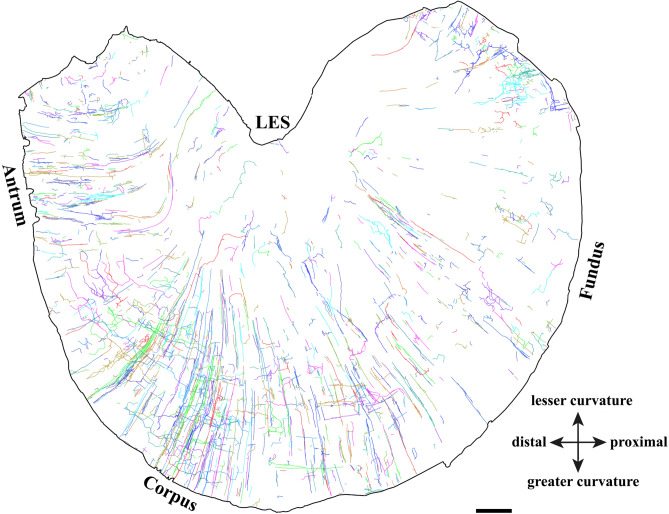
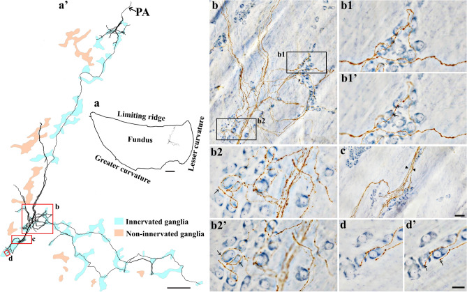
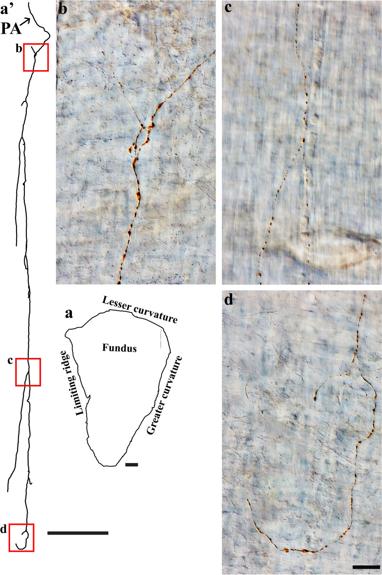
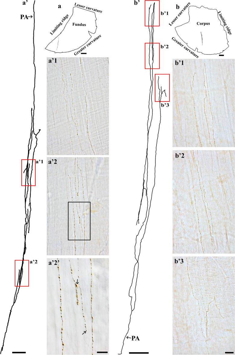
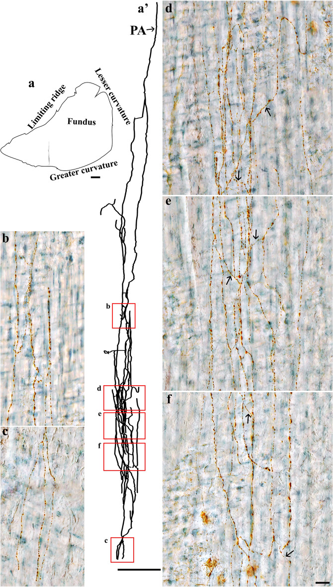
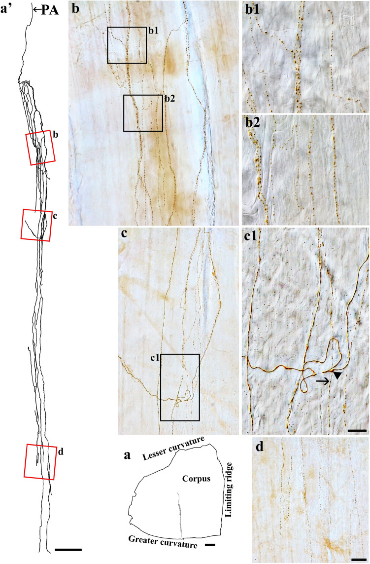
The dorsal root ganglia (DRG) project spinal afferent axons to the stomach. However, the distribution and morphology of spinal afferent axons in the stomach have not been well characterized. In this study, we used a combination of state-of-the-art techniques, including anterograde tracer injection into the left DRG T7-T11, avidin-biotin and Cuprolinic Blue labeling, Zeiss M2 Imager, and Neurolucida to characterize spinal afferent axons in flat-mounts of the whole rat stomach muscular wall. We found that spinal afferent axons innervated all regions with a variety of distinct terminal structures innervating different gastric targets: (1) The ganglionic type: some axons formed varicose contacts with individual neurons within myenteric ganglia. (2) The muscle type: most axons ran in parallel with the longitudinal and circular muscles and expressed spherical varicosities. Complex terminal structures were observed within the circular muscle layer. (3) The ganglia-muscle mixed type: some individual varicose axons innervated both myenteric neurons and the circular muscle, exhibiting polymorphic terminal structures. (4) The vascular type: individual varicose axons ran along the blood vessels and occasionally traversed the vessel wall. This work provides a foundation for future topographical anatomical and functional mapping of spinal afferent axon innervation of the stomach under normal and pathophysiological conditions.
背根神经节 (DRG) 将脊髓传入轴突投射到胃。然而,脊髓传入轴突在胃中的分布和形态尚未得到很好的描述。在这项研究中,我们使用了一系列先进技术,包括将示踪剂注入左侧 DRG T7-T11,亲和素-生物素和 Cuprolinic Blue 标记、蔡司 M2 Imager 和 Neurolucida,以描绘整个大鼠胃壁平滑肌层的脊髓传入轴突。我们发现,脊髓传入轴突支配所有区域,具有多种不同的终末结构,支配不同的胃靶标:(1)神经节型:一些轴突与肠神经节内的单个神经元形成曲张接触。(2)肌肉型:大多数轴突与纵肌和环肌平行运行,并表达球形曲张体。在环肌层内观察到复杂的终末结构。(3)神经节-肌肉混合型:一些个体曲张轴突同时支配肠神经元和环肌,表现出多态性终末结构。(4)血管型:个别曲张轴突沿血管运行,偶尔穿过血管壁。这项工作为未来在正常和病理生理条件下对胃的脊髓传入轴突支配进行拓扑解剖和功能映射奠定了基础。