Department of Biology, Rosenstiel Basic Medical Science Research Center, Brandeis University, Waltham, Massachusetts, USA.
Department of Biochemistry and Molecular Biology, Drexel University, Philadelphia, Pennsylvania, USA.
J Biol Chem. 2023 Dec;299(12):105367. doi: 10.1016/j.jbc.2023.105367. Epub 2023 Oct 19.
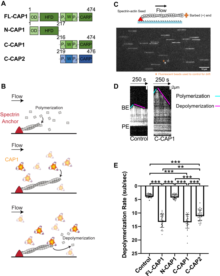
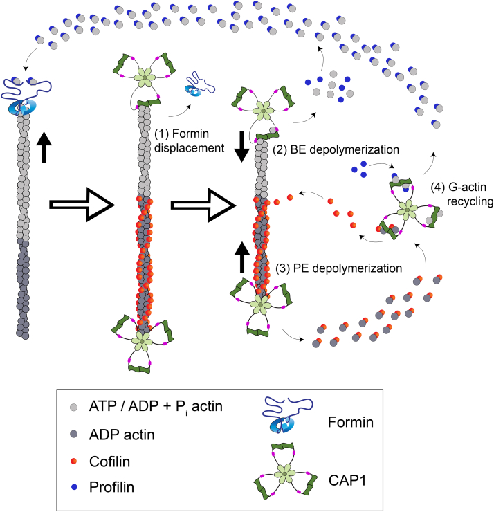
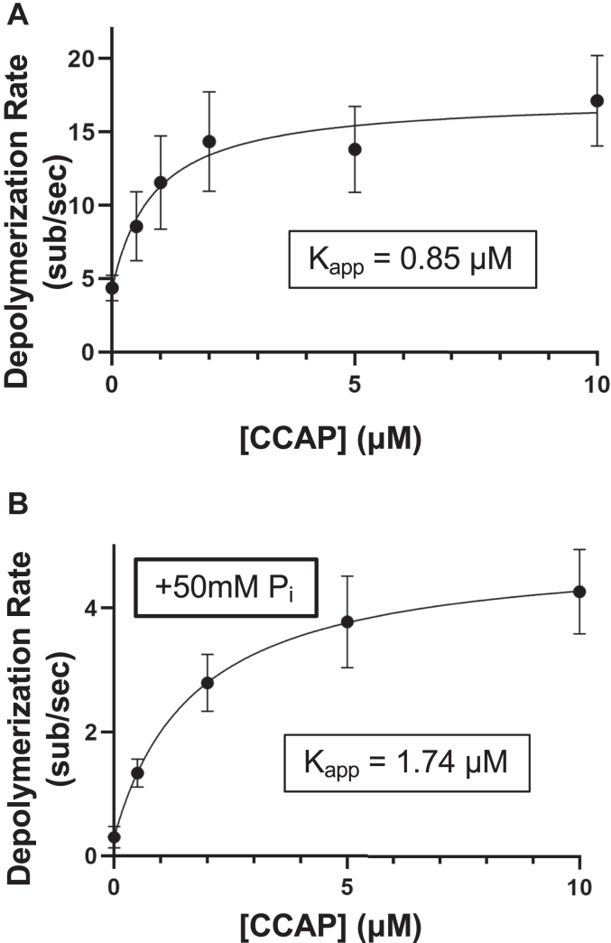
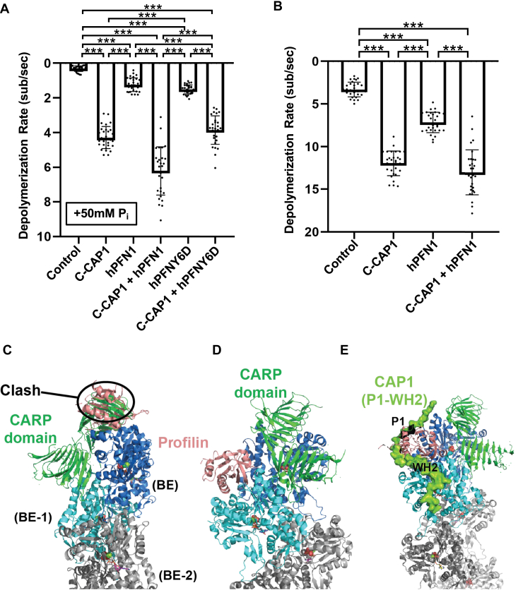
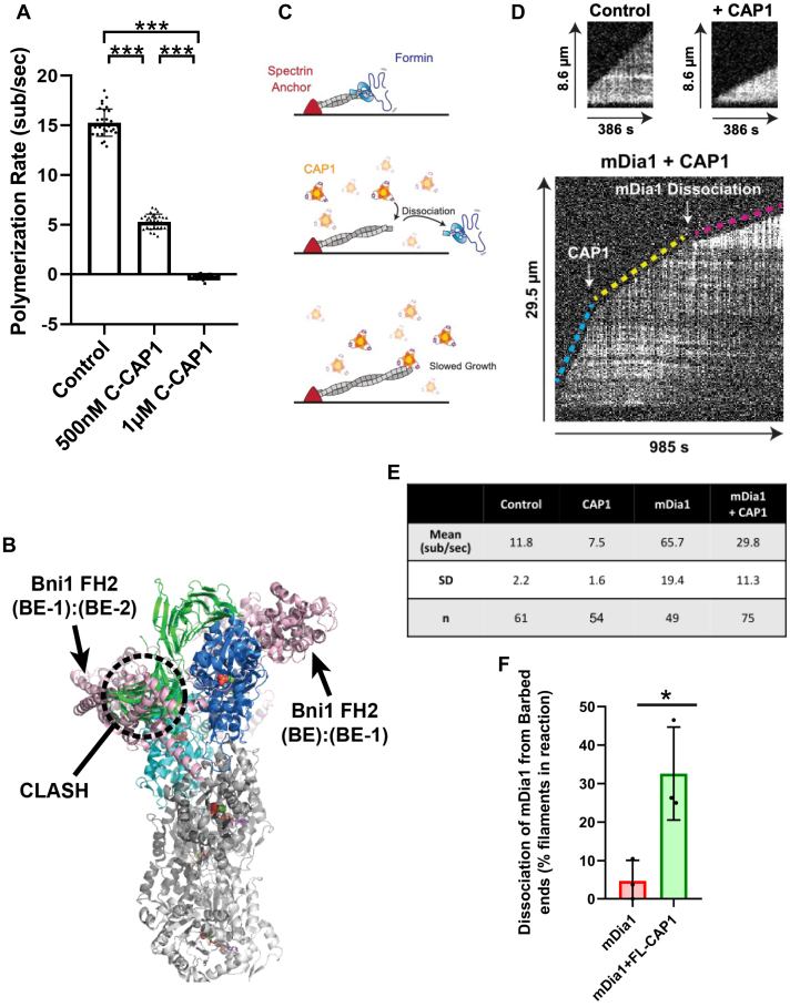
Cyclase-associated protein (CAP) has emerged as a central player in cellular actin turnover, but its molecular mechanisms of action are not yet fully understood. Recent studies revealed that the N terminus of CAP interacts with the pointed ends of actin filaments to accelerate depolymerization in conjunction with cofilin. Here, we use in vitro microfluidics-assisted TIRF microscopy to show that the C terminus of CAP promotes depolymerization at the opposite (barbed) ends of actin filaments. In the absence of actin monomers, full-length mouse CAP1 and C-terminal halves of CAP1 (C-CAP1) and CAP2 (C-CAP2) accelerate barbed end depolymerization. Using mutagenesis and structural modeling, we show that these activities are mediated by the WH2 and CARP domains of CAP. In addition, we observe that CAP collaborates with profilin to accelerate barbed end depolymerization and that these effects depend on their direct interaction, providing the first known example of CAP-profilin collaborative effects in regulating actin. In the presence of actin monomers, CAP1 attenuates barbed end growth and promotes formin dissociation. Overall, these findings demonstrate that CAP uses distinct domains and mechanisms to interact with opposite ends of actin filaments and drive turnover. Further, they contribute to the emerging view of actin barbed ends as sites of dynamic molecular regulation, where numerous proteins compete and cooperate with each other to tune polymer dynamics, similar to the rich complexity seen at microtubule ends.
环化酶相关蛋白(CAP)已成为细胞肌动蛋白周转的核心参与者,但它的作用机制尚不完全清楚。最近的研究表明,CAP 的 N 端与肌动蛋白丝的尖端相互作用,与丝切蛋白一起加速解聚。在这里,我们使用体外微流控辅助 TIRF 显微镜显示 CAP 的 C 端促进肌动蛋白丝的反(棘)端解聚。在没有肌动蛋白单体的情况下,全长小鼠 CAP1 和 CAP1(C-CAP1)和 CAP2(C-CAP2)的 C 端促进棘突端解聚。通过突变和结构建模,我们表明这些活性是由 CAP 的 WH2 和 CARP 结构域介导的。此外,我们观察到 CAP 与 Profilin 协同作用以加速棘突端解聚,并且这些效应取决于它们的直接相互作用,为 CAP-Profilin 在调节肌动蛋白方面的协同作用提供了第一个已知实例。在肌动蛋白单体存在的情况下,CAP1 减弱了棘突端的生长并促进了形成素的解离。总的来说,这些发现表明 CAP 使用不同的结构域和机制与肌动蛋白丝的相反末端相互作用并驱动周转率。此外,它们有助于新兴的肌动蛋白棘突作为动态分子调节的位点的观点,其中许多蛋白质相互竞争和合作以调节聚合物动力学,类似于在微管末端看到的丰富复杂性。