Taghian Dinani Hengameh, Naderi Nushin, Tavalaee Marziyeh, Rabiee Farzaneh, Nasr-Esfahani Mohammad Hossein
ACECR Institute of Higher Education, Isfahan Branch, Isfahan, Iran.
Department of Animal Biotechnology, Reproductive Biomedicine Research Center, Royan Institute for Biotechnology, ACECR, Isfahan, Iran.
Cell J. 2023 Oct 9;25(10):706-716. doi: 10.22074/cellj.2023.2000170.1284.
Epigenetic modifications such as DNA methylation play a key role in male infertility etiology. This study aimed to explore the global DNA methylation status in testicular spermatogenic cells of varicocele-induced rats and consider their semen quality, with a focus on key epigenetic marks, namely 5-methylcytosine (5-mC) and 5-hydroxymethylcytosine (5-hmC), as well as the mRNA and proteins of ten-eleven translocation (TET) methylcytosine dioxygenases 1-3.
In this experimental study, 24 mature male Wistar rats (8 in each group) were assigned amongst the control, sham, and varicocele groups. Sperm quality was assessed, and DNA methylation patterns of testicular spermatogenic cells were investigated using reverse transcription-polymerase chain reaction (RT-PCR), western blot, and immunofluorescence techniques.
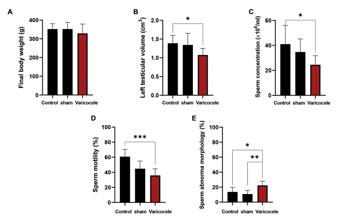
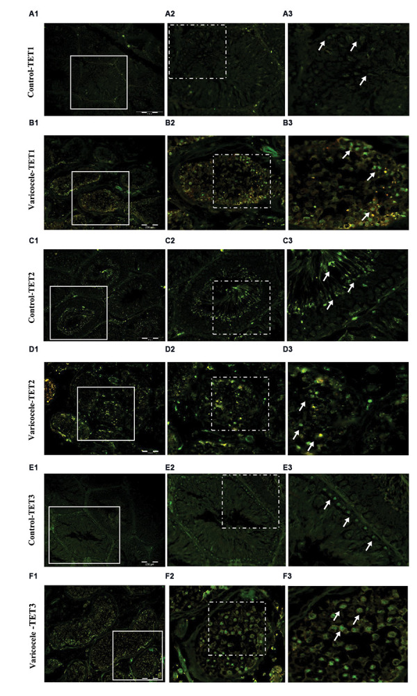
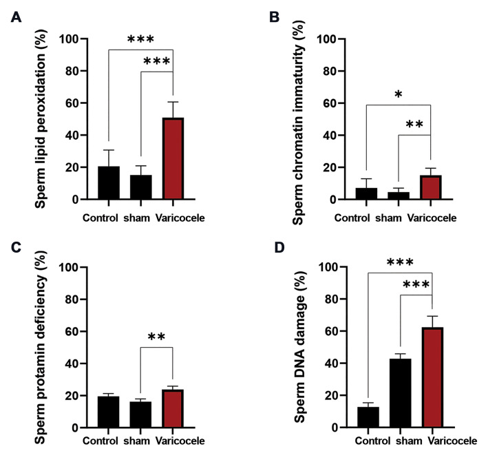
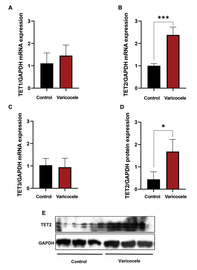
Sperm parameters, chromatin and DNA integrity were significantly lower, and sperm lipid peroxidation significantly increased in varicocele-induced rats in comparison with control rats. During spermatogenesis in rat testis, 5-mC and 5-hmC epigenetic marks, and TET1-3 mRNA and proteins were expressed. In contrast to the 5-mC fluorescent signal which was presented in all testicular cells, the 5-hmC fluorescent signal was presented exclusively in spermatogonia and a few spermatids. In varicocele-induced rats, the 5-mC signal decreased in all cells within the tubules, whereas a strong signal of 5-hmC was detected in seminiferous tubules compared to the control group. As well, the levels of TET2 mRNA and protein expression were significantly upregulated in varicocele-induced rats in comparison with the control group. Also, our results showed that the varicocele-induced animals exhibited strong fluorescent signals of TET1-3 in testicular cells, whereas weak fluorescent signals were identified in the seminiferous tubules of the control animals.
Consequently, we showed TET2 upregulation and the 5-hmC gain at testicular levels are associated with varicocele and sperm quality decline, and therefore they can be exploited as potential biomarkers of spermatogenesis.
DNA甲基化等表观遗传修饰在男性不育病因中起关键作用。本研究旨在探讨精索静脉曲张诱导大鼠睾丸生精细胞中的整体DNA甲基化状态,并考量其精液质量,重点关注关键表观遗传标记,即5-甲基胞嘧啶(5-mC)和5-羟甲基胞嘧啶(5-hmC),以及11-易位(TET)甲基胞嘧啶双加氧酶1-3的mRNA和蛋白质。
在本实验研究中,24只成熟雄性Wistar大鼠(每组8只)被分配至对照组、假手术组和精索静脉曲张组。评估精子质量,并使用逆转录聚合酶链反应(RT-PCR)、蛋白质免疫印迹和免疫荧光技术研究睾丸生精细胞的DNA甲基化模式。
与对照大鼠相比,精索静脉曲张诱导的大鼠精子参数、染色质和DNA完整性显著降低,精子脂质过氧化显著增加。在大鼠睾丸生精过程中,5-mC和5-hmC表观遗传标记以及TET1-3 mRNA和蛋白质均有表达。与在所有睾丸细胞中呈现的5-mC荧光信号不同,5-hmC荧光信号仅出现在精原细胞和少数精子细胞中。在精索静脉曲张诱导的大鼠中,小管内所有细胞中的5-mC信号均降低,而与对照组相比,在生精小管中检测到强烈的5-hmC信号。此外,与对照组相比,精索静脉曲张诱导的大鼠中TET2 mRNA和蛋白质表达水平显著上调。而且,我们的结果表明,精索静脉曲张诱导的动物在睾丸细胞中呈现TET1-3的强荧光信号,而在对照动物的生精小管中鉴定出弱荧光信号。
因此,我们表明睾丸水平上TET2上调和5-hmC增加与精索静脉曲张和精子质量下降相关,因此它们可被用作生精的潜在生物标志物。