Shin Sang Hwa, Cha Yoon Ki, Kim Jong Hee, Woo Jung Han, Kim Tae Jung, Chung Myung Jin, Han Joungho, Lim Yun-Jung
Department of Radiology and Center for Imaging Science, Samsung Medical Center, Sungkyunkwan University School of Medicine, Seoul, Republic of Korea.
Department of Pathology, Samsung Medical Center, Sungkyunkwan University School of Medicine, Seoul, Republic of Korea.
J Thorac Dis. 2023 Sep 28;15(9):4818-4825. doi: 10.21037/jtd-23-733. Epub 2023 Aug 25.
Placental transmogrification of the lung is a very rare benign lung disease with a characteristic finding being alveoli resembling chorionic villi of the placenta. The purpose of this study was to assess the computed tomography (CT) findings of placental transmogrification of the lung in six patients and their relation to the histopathologic findings.
Six patients with histopathologically proven placental transmogrification of the lung from 2004 to 2021 were included. Their CT findings were analyzed and their imaging features were compared with pathology specimens.
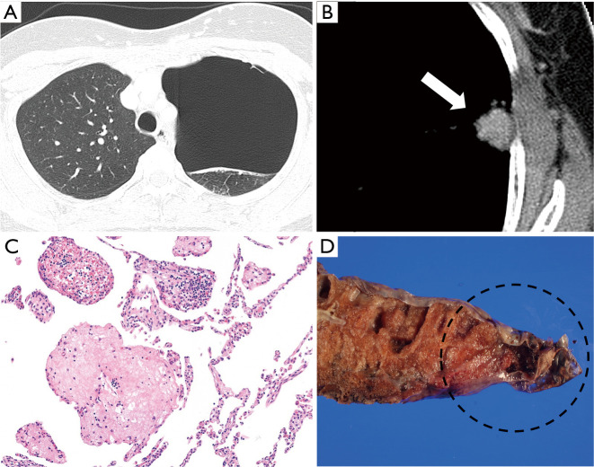
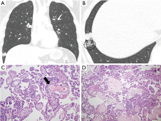
In four of six cases, CT showed variable sized cystic lesions confined to a unilateral lung. One case presented nodule and cystic lesion together. The other case showed solitary pulmonary nodule without cystic lesion. Moreover, nodular interlobular septal thickening and clustered interstitial nodules were observed in all six cases. In four of the six cases, these nodules merged into dense nodular consolidation. Three cases showed dilated pulmonary vasculatures of the involved lung.
On CT, placental transmogrification of the lung typically presents as cystic lesion confined to a unilateral lung. Pulmonary nodule with or without associated cystic lesion can also be seen. Nodular interlobular septal thickening and clustered interstitial nodules were observed in all cases. This might be attributable to the proliferation of chorionic villi-like structures in interstitium which are found in histopathologic specimens.
肺胎盘样化生是一种非常罕见的良性肺部疾病,其特征性表现为肺泡类似胎盘的绒毛膜绒毛。本研究的目的是评估6例肺胎盘样化生患者的计算机断层扫描(CT)表现及其与组织病理学结果的关系。
纳入2004年至2021年间6例经组织病理学证实为肺胎盘样化生的患者。分析其CT表现,并将其影像学特征与病理标本进行比较。
6例中有4例CT显示大小不一的囊性病变局限于单侧肺。1例同时出现结节和囊性病变。另1例表现为孤立性肺结节,无囊性病变。此外,所有6例均观察到结节状小叶间隔增厚和簇状间质结节。6例中有4例,这些结节融合成致密的结节状实变。3例显示受累肺的肺血管扩张。
在CT上,肺胎盘样化生通常表现为局限于单侧肺的囊性病变。也可见伴有或不伴有相关囊性病变的肺结节。所有病例均观察到结节状小叶间隔增厚和簇状间质结节。这可能归因于组织病理学标本中发现的间质中绒毛膜绒毛样结构的增殖。