Welsh Ian C, Feiler Maria E, Lipman Danika, Mormile Isabel, Hansen Karissa, Percival Christopher J
Program in Craniofacial Biology, University of California at San Francisco, San Francisco, California 94143, USA.
Department of Orofacial Sciences, University of California at San Francisco, San Francisco, California 94143, USA.
bioRxiv. 2024 Nov 13:2023.10.03.560703. doi: 10.1101/2023.10.03.560703.
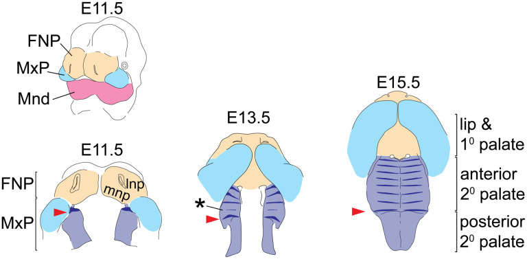
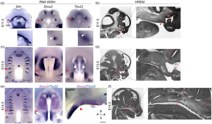
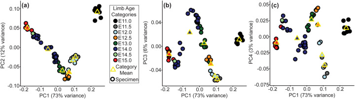
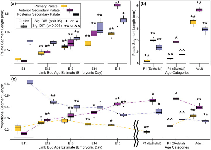
Anterior-posterior (A-P) elongation of the palate is a critical aspect of integrated midfacial morphogenesis. Reciprocal epithelial-mesenchymal interactions drive secondary palate elongation that is coupled to the periodic formation of signaling centers within the rugae growth zone (RGZ). However, the relationship between RGZ-driven morphogenetic processes, the differentiative dynamics of underlying palatal bone mesenchymal precursors, and the segmental organization of the upper jaw has remained enigmatic. A detailed ontogenetic study of these relationships is important because palatal segment growth is a critical aspect of normal midfacial growth, can produce dysmorphology when altered, and is a likely basis for evolutionary differences in upper jaw morphology. We completed a combined whole mount gene expression and morphometric analysis of normal murine palatal segment growth dynamics and resulting upper jaw morphology. Our results demonstrated that the first formed palatal ruga (ruga 1), found just posterior to the RGZ, maintained an association with important nasal, neurovascular and palatal structures throughout early midfacial development. This suggested that these features are positioned at a proximal source of embryonic midfacial directional growth. Our detailed characterization of midfacial morphogenesis revealed a one-to-one relationship between palatal segments and upper jaw bones during the earliest stages of palatal elongation. Growth of the maxillary anlage within the anterior secondary palate is uniquely coupled to RGZ-driven morphogenesis. This may help drive the unequaled proportional elongation of the anterior secondary palate segment prior to palatal shelf fusion. Our results also demonstrated that the future maxillary-palatine suture, approximated by the position of ruga 1 and consistently associated with the palatine anlage, formed predominantly via the posterior differentiation of the maxilla within the expanding anterior secondary palate. Our ontogenetic analysis provides a novel and detailed picture of the earliest spatiotemporal dynamics of intramembranous midfacial skeletal specification and differentiation within the context of the surrounding palatal segment AP elongation and associated rugae formation.
腭的前后(A-P)伸长是中面部整体形态发生的一个关键方面。上皮-间充质的相互作用驱动继发腭的伸长,这与嵴生长区(RGZ)内信号中心的周期性形成相关。然而,RGZ驱动的形态发生过程、腭骨间充质前体的分化动力学以及上颌的节段组织之间的关系仍然不明。对这些关系进行详细的个体发生学研究很重要,因为腭节段生长是正常中面部生长的一个关键方面,改变时会产生畸形,并且可能是上颌形态进化差异的一个基础。我们完成了对正常小鼠腭节段生长动力学及由此产生的上颌形态的全组织基因表达和形态测量分析的联合研究。我们的结果表明,最早形成的腭嵴(嵴1)位于RGZ后方,在整个早期中面部发育过程中与重要的鼻腔、神经血管和腭结构保持关联。这表明这些特征位于胚胎中面部定向生长的近端来源处。我们对中面部形态发生的详细表征揭示了在腭伸长的最早阶段,腭节段与上颌骨之间存在一对一的关系。前继发腭内上颌原基的生长与RGZ驱动的形态发生独特地相关联。这可能有助于在腭板融合之前驱动前继发腭节段不成比例的伸长。我们的结果还表明,由嵴1的位置近似并始终与腭原基相关联的未来上颌-腭缝,主要通过扩展的前继发腭内上颌骨的后部分化形成。我们的个体发生学分析提供了一幅新颖而详细的膜内中面部骨骼特化和分化的最早时空动态图,该动态图处于周围腭节段AP伸长及相关嵴形成的背景下。