Institute of Biological Information Processing, Structural Biochemistry (IBI-7), Forschungszentrum Jülich, Jülich, Germany.
JuStruct, Jülich Center for Structural Biology, Forschungszentrum Jülich, Jülich, Germany.
Nat Neurosci. 2023 Dec;26(12):2073-2080. doi: 10.1038/s41593-023-01484-4. Epub 2023 Nov 16.
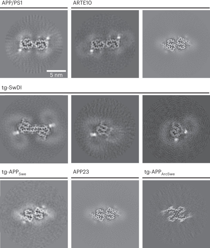
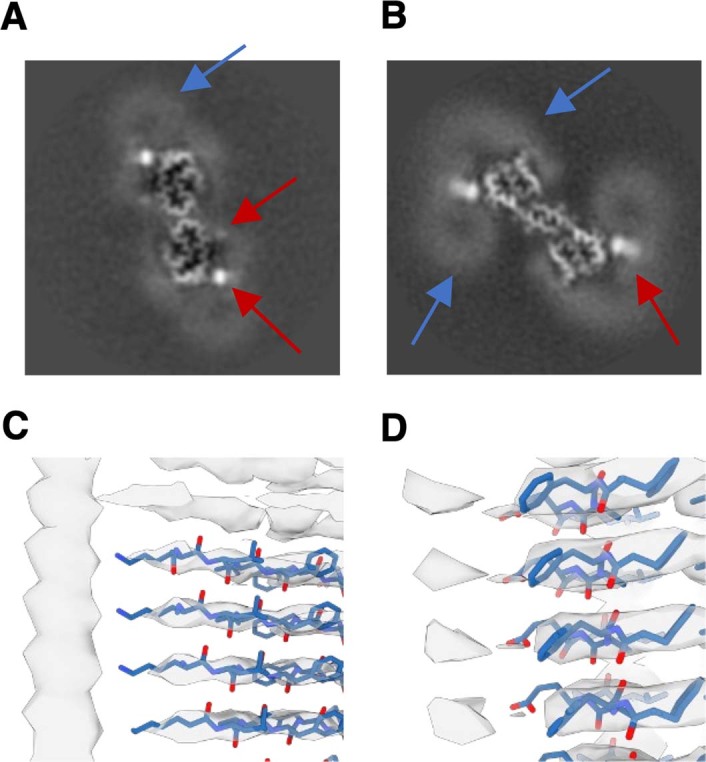
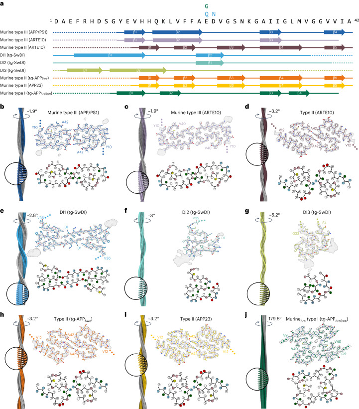
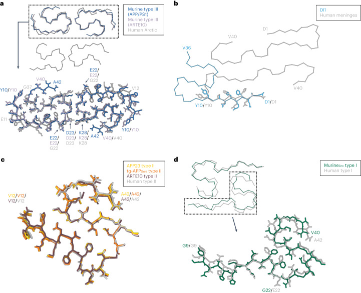
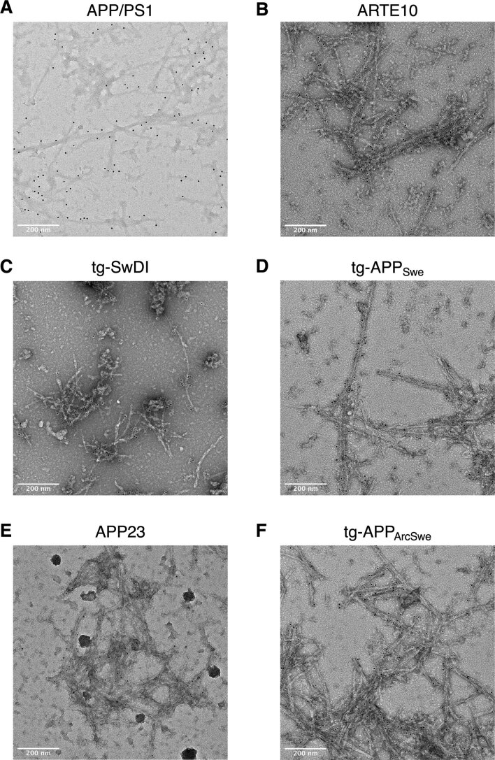
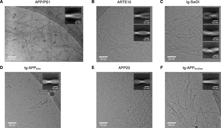
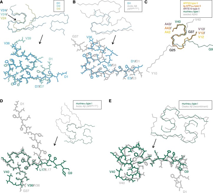
The use of transgenic mice displaying amyloid-β (Aβ) brain pathology has been essential for the preclinical assessment of new treatment strategies for Alzheimer's disease. However, the properties of Aβ in such mice have not been systematically compared to Aβ in the brains of patients with Alzheimer's disease. Here, we determined the structures of nine ex vivo Aβ fibrils from six different mouse models by cryogenic-electron microscopy. We found novel Aβ fibril structures in the APP/PS1, ARTE10 and tg-SwDI models, whereas the human type II filament fold was found in the ARTE10, tg-APP and APP23 models. The tg-APP mice showed an Aβ fibril whose structure resembles the human type I filament found in patients with sporadic Alzheimer's disease. A detailed assessment of the Aβ fibril structure is key to the selection of adequate mouse models for the preclinical development of novel plaque-targeting therapeutics and positron emission tomography imaging tracers in Alzheimer's disease.
使用显示淀粉样蛋白-β(Aβ)脑病理学的转基因小鼠对于评估阿尔茨海默病新治疗策略的临床前评估至关重要。然而,此类小鼠中 Aβ的特性尚未与阿尔茨海默病患者脑中的 Aβ进行系统比较。在这里,我们通过低温电子显微镜确定了来自六个不同小鼠模型的九种体外 Aβ纤维的结构。我们在 APP/PS1、ARTE10 和 tg-SwDI 模型中发现了新型 Aβ纤维结构,而在 ARTE10、tg-APP 和 APP23 模型中发现了人类 II 型丝折叠。Tg-APP 小鼠显示出一种 Aβ纤维,其结构类似于在散发性阿尔茨海默病患者中发现的人类 I 型丝。对 Aβ纤维结构的详细评估是选择合适的小鼠模型进行新型斑块靶向治疗药物和正电子发射断层扫描(PET)成像示踪剂在阿尔茨海默病中的临床前开发的关键。