NHC Key Laboratory of Birth Defect for Research and Prevention, Hunan Provincial Maternal and Child Health Care Hospital, Changsha, Hunan, China.
Department of Epidemiology and Health Statistics, Xiangya School of Public Health, Central South University, Changsha, Hunan, China.
Stem Cell Res Ther. 2023 Nov 19;14(1):336. doi: 10.1186/s13287-023-03528-9.
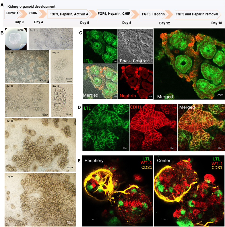
Kidney organoids derived from human pluripotent stem cells (HiPSCs) hold huge applications for drug screening, disease modeling, and cell transplanting therapy. However, these applications are limited since kidney organoid cannot maintain complete morphology and function like human kidney. Kidney organoids are not well differentiated since the core of the organoid lacked oxygen, nutrition, and vasculature, which creates essential niches. Hypoxia-inducible factor-1 α (HIF-1α) serves as a critical regulator in vascularization and cell survival under hypoxia environment. Less is known about the role of HIF-1α in kidney organoids in this regard. This study tried to investigate the effect of HIF-1α in kidney organoid vascularization and related disease modeling.
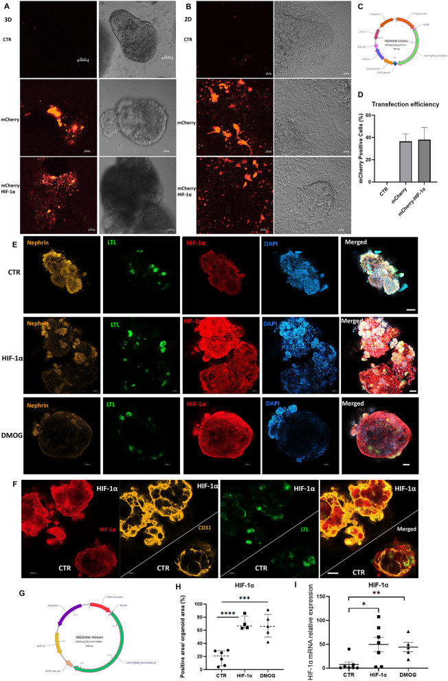
For the vascularization study, kidney organoids were generated from human induced pluripotent stem cells. We overexpressed HIF-1α via plasmid transfection or treated DMOG (Dimethyloxallyl Glycine, an agent for HIF-1α stabilization and accumulation) in kidney progenitor cells to detect the endothelium. For the disease modeling study, we treated kidney organoid with cisplatin under hypoxia environment, with additional HIF-1α transfection.
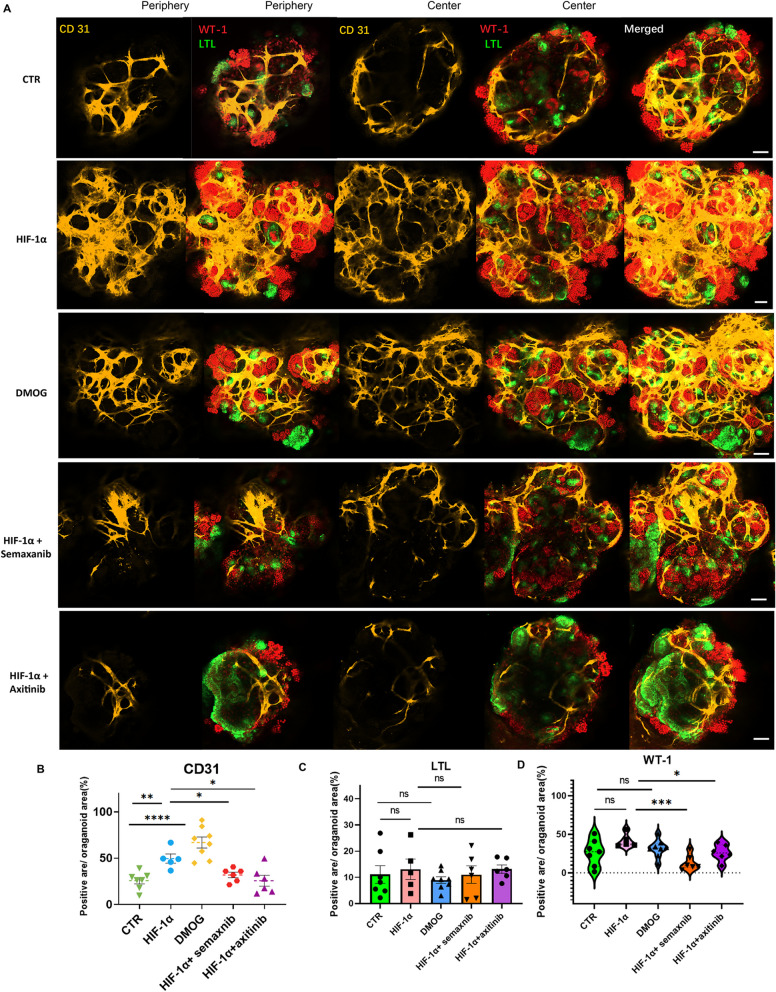
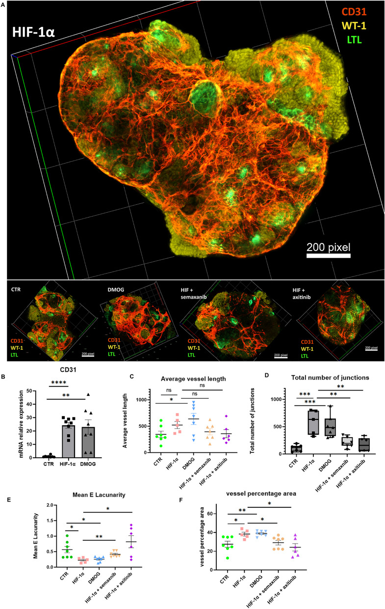
HIF-1α overexpression elicited kidney organoid vascularization. The endothelial cells and angiotool analysis parameters were increased in HIF-1α plasmid-transfected and DMOG-treated organoids. These angiogenesis processes were partially blocked by VEGFR inhibitors, semaxanib or axitinib. Cisplatin-induced kidney injury (Cleaved caspase 3) was protected by HIF-1α through the upregulation of CD31 and SOD2.
We demonstrated that HIF-1α elicited the process of kidney organoid vascularization and protected against cisplatin-induced kidney organoid injury in hypoxia environment.
来源于人类多能干细胞(HiPSCs)的肾脏类器官在药物筛选、疾病建模和细胞移植治疗方面具有巨大的应用潜力。然而,由于肾脏类器官无法像人类肾脏那样保持完整的形态和功能,这些应用受到了限制。由于类器官的核心缺乏氧气、营养和血管,因此无法很好地分化,这就创造了必要的生态位。缺氧诱导因子-1α(HIF-1α)在缺氧环境下的血管生成和细胞存活中起着关键的调节作用。关于 HIF-1α在肾脏类器官中的作用,人们知之甚少。本研究试图探讨 HIF-1α在肾脏类器官血管生成及相关疾病建模中的作用。
为了进行血管生成研究,我们从人诱导多能干细胞中生成肾脏类器官。我们通过质粒转染过表达 HIF-1α,或用 DMOG(二甲氧乙基亚硝氨酸,一种稳定和积累 HIF-1α 的试剂)处理肾祖细胞,以检测内皮细胞。在疾病建模研究中,我们在缺氧环境下用顺铂处理肾脏类器官,并进行 HIF-1α 转染。
HIF-1α 的过表达引发了肾脏类器官的血管生成。在 HIF-1α 质粒转染和 DMOG 处理的类器官中,内皮细胞和血管生成分析参数增加。这些血管生成过程被 VEGFR 抑制剂,semaxanib 或 axitinib 部分阻断。HIF-1α 通过上调 CD31 和 SOD2 来保护顺铂诱导的肾脏类器官损伤(Cleaved caspase 3)。
我们证明了 HIF-1α 引发了肾脏类器官的血管生成过程,并在缺氧环境下保护顺铂诱导的肾脏类器官损伤。