• 文献检索
  • 文档翻译
  • 深度研究
  • 学术资讯
  • Suppr Zotero 插件Zotero 插件
  • 邀请有礼
  • 套餐&价格
  • 历史记录
应用&插件
Suppr Zotero 插件Zotero 插件浏览器插件Mac 客户端Windows 客户端微信小程序
定价
高级版会员购买积分包购买API积分包
服务
文献检索文档翻译深度研究API 文档MCP 服务
关于我们
关于 Suppr公司介绍联系我们用户协议隐私条款
关注我们

Suppr 超能文献

核心技术专利:CN118964589B侵权必究
粤ICP备2023148730 号-1Suppr @ 2026

文献检索

告别复杂PubMed语法,用中文像聊天一样搜索,搜遍4000万医学文献。AI智能推荐,让科研检索更轻松。

立即免费搜索

文件翻译

保留排版,准确专业,支持PDF/Word/PPT等文件格式,支持 12+语言互译。

免费翻译文档

深度研究

AI帮你快速写综述,25分钟生成高质量综述,智能提取关键信息,辅助科研写作。

立即免费体验

基于图神经网络的乳腺癌诊断,使用超声图像,并通过整合具有医学意义的特征的优化图构建进行。

Graph neural network-based breast cancer diagnosis using ultrasound images with optimized graph construction integrating the medically significant features.

机构信息

Faculty of Science and Technology, Charles Darwin University, Casuarina, NT, 0909, Australia.

Health Informatics Research Laboratory (HIRL), Department of Computer Science and Engineering, Daffodil International University, Dhaka, 1216, Bangladesh.

出版信息

J Cancer Res Clin Oncol. 2023 Dec;149(20):18039-18064. doi: 10.1007/s00432-023-05464-w. Epub 2023 Nov 20.

DOI:10.1007/s00432-023-05464-w
PMID:37982829
原文链接:https://pmc.ncbi.nlm.nih.gov/articles/PMC10725367/
Abstract

PURPOSE

An automated computerized approach can aid radiologists in the early diagnosis of breast cancer. In this study, a novel method is proposed for classifying breast tumors into benign and malignant, based on the ultrasound images through a Graph Neural Network (GNN) model utilizing clinically significant features.

METHOD

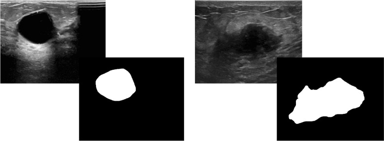
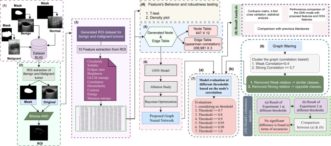
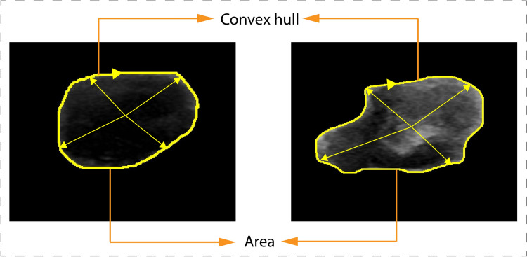
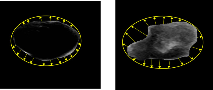
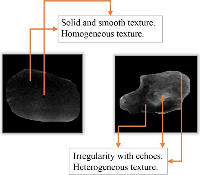
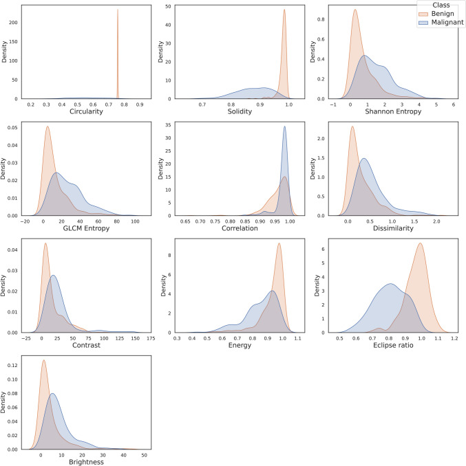
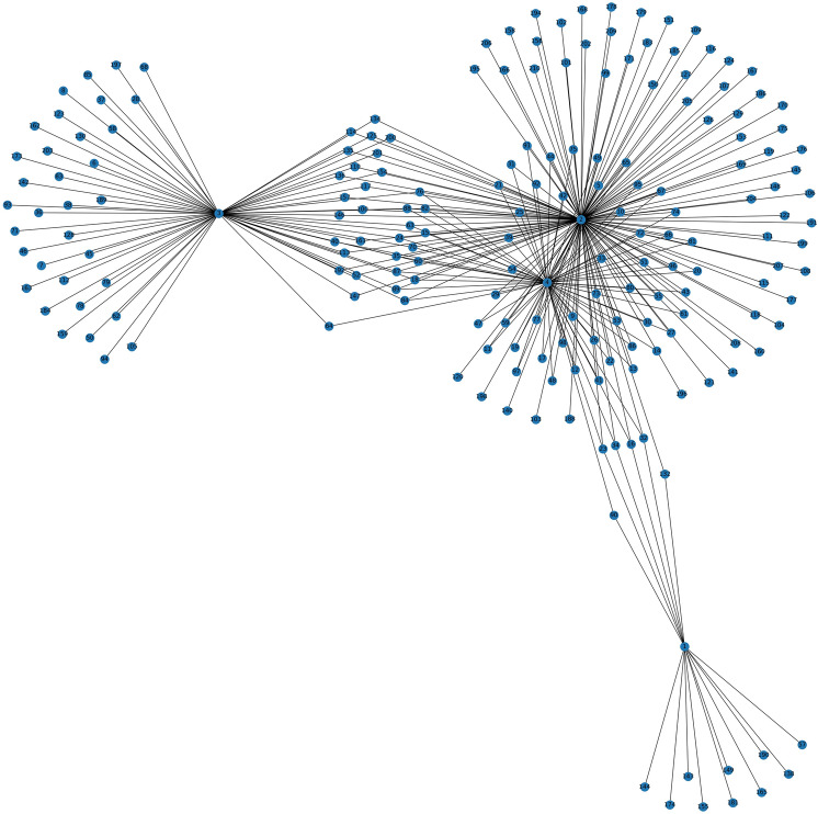
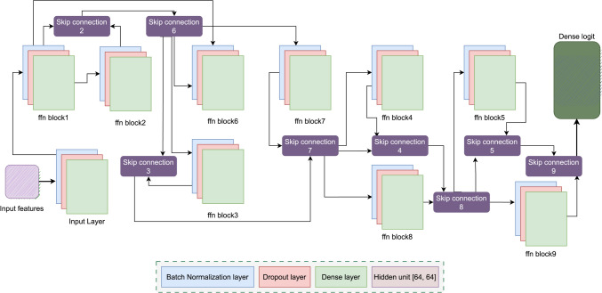
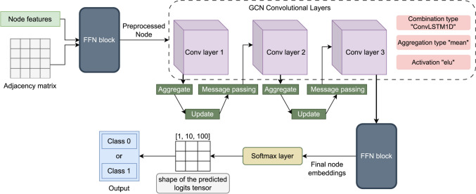
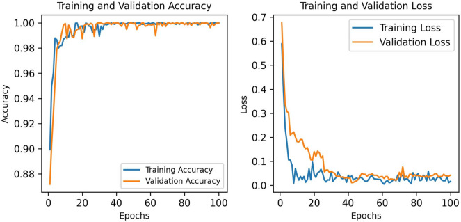
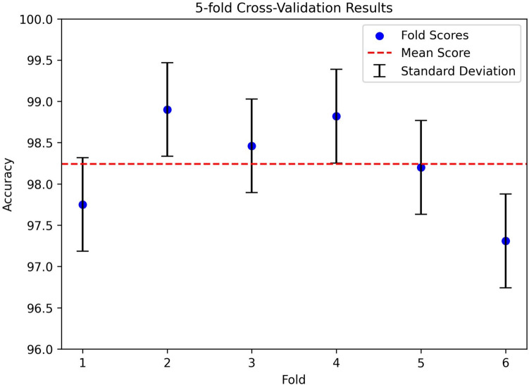
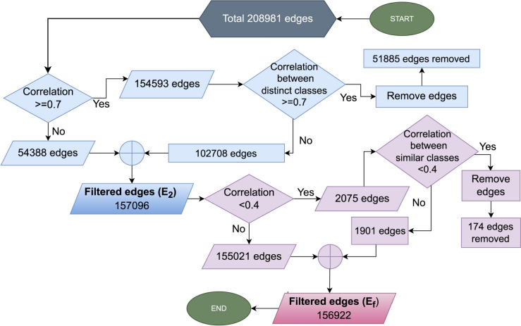
Ten informative features are extracted from the region of interest (ROI), based on the radiologists' diagnosis markers. The significance of the features is evaluated using density plot and T test statistical analysis method. A feature table is generated where each row represents individual image, considered as node, and the edges between the nodes are denoted by calculating the Spearman correlation coefficient. A graph dataset is generated and fed into the GNN model. The model is configured through ablation study and Bayesian optimization. The optimized model is then evaluated with different correlation thresholds for getting the highest performance with a shallow graph. The performance consistency is validated with k-fold cross validation. The impact of utilizing ROIs and handcrafted features for breast tumor classification is evaluated by comparing the model's performance with Histogram of Oriented Gradients (HOG) descriptor features from the entire ultrasound image. Lastly, a clustering-based analysis is performed to generate a new filtered graph, considering weak and strong relationships of the nodes, based on the similarities.

RESULTS

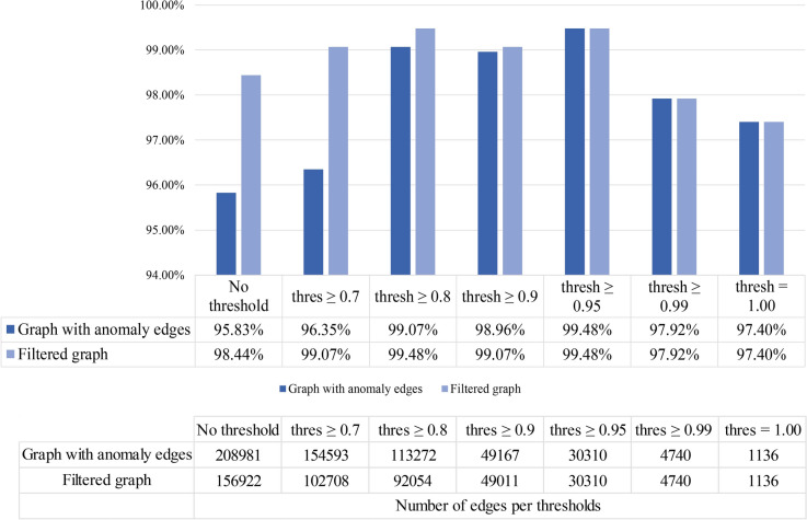
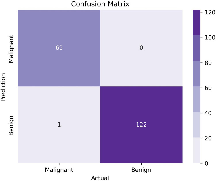
The results indicate that with a threshold value of 0.95, the GNN model achieves the highest test accuracy of 99.48%, precision and recall of 100%, and F1 score of 99.28%, reducing the number of edges by 85.5%. The GNN model's performance is 86.91%, considering no threshold value for the graph generated from HOG descriptor features. Different threshold values for the Spearman's correlation score are experimented with and the performance is compared. No significant differences are observed between the previous graph and the filtered graph.

CONCLUSION

The proposed approach might aid the radiologists in effective diagnosing and learning tumor pattern of breast cancer.

摘要

目的

自动化计算机方法可以帮助放射科医生早期诊断乳腺癌。在这项研究中,提出了一种基于图形神经网络(GNN)模型的新方法,该方法利用临床有意义的特征对乳腺肿瘤进行良性和恶性分类。

方法

从感兴趣区域(ROI)中提取 10 个信息特征,基于放射科医生的诊断标记。使用密度图和 T 检验统计分析方法评估特征的显著性。生成一个特征表,其中每一行代表一个单独的图像,视为节点,节点之间的边由计算 Spearman 相关系数来表示。生成一个图数据集,并将其输入到 GNN 模型中。通过消融研究和贝叶斯优化来配置模型。然后,使用不同的相关阈值来评估优化后的模型,以获得浅图的最高性能。通过 k 折交叉验证验证性能一致性。通过将模型的性能与整个超声图像的方向梯度直方图(HOG)描述符特征进行比较,评估利用 ROI 和手工制作特征进行乳腺肿瘤分类的影响。最后,根据相似性,基于节点的弱和强关系,进行基于聚类的分析,生成一个新的过滤图。

结果

结果表明,在阈值为 0.95 的情况下,GNN 模型的测试准确率最高为 99.48%,精度和召回率均为 100%,F1 评分为 99.28%,减少了 85.5%的边数。考虑到生成的 HOG 描述符特征的图没有阈值,GNN 模型的性能为 86.91%。对 Spearman 相关得分的不同阈值进行实验,并比较性能。未观察到前一个图和过滤图之间有显著差异。

结论

该方法可能有助于放射科医生有效诊断和学习乳腺癌肿瘤模式。

https://cdn.ncbi.nlm.nih.gov/pmc/blobs/7855/11797365/fe9fa7ed4a3b/432_2023_5464_Fig15_HTML.jpg
https://cdn.ncbi.nlm.nih.gov/pmc/blobs/7855/11797365/00daaf4a9055/432_2023_5464_Fig1_HTML.jpg
https://cdn.ncbi.nlm.nih.gov/pmc/blobs/7855/11797365/bf5f1c3b2d70/432_2023_5464_Fig2_HTML.jpg
https://cdn.ncbi.nlm.nih.gov/pmc/blobs/7855/11797365/f195677c9a3c/432_2023_5464_Fig3_HTML.jpg
https://cdn.ncbi.nlm.nih.gov/pmc/blobs/7855/11797365/bb5e841e6284/432_2023_5464_Fig4_HTML.jpg
https://cdn.ncbi.nlm.nih.gov/pmc/blobs/7855/11797365/21391315a740/432_2023_5464_Fig5_HTML.jpg
https://cdn.ncbi.nlm.nih.gov/pmc/blobs/7855/11797365/b91138b1d27f/432_2023_5464_Fig6_HTML.jpg
https://cdn.ncbi.nlm.nih.gov/pmc/blobs/7855/11797365/e98177249d2b/432_2023_5464_Fig7_HTML.jpg
https://cdn.ncbi.nlm.nih.gov/pmc/blobs/7855/11797365/58c27a9784c5/432_2023_5464_Fig8_HTML.jpg
https://cdn.ncbi.nlm.nih.gov/pmc/blobs/7855/11797365/b8ab699ecc29/432_2023_5464_Fig9_HTML.jpg
https://cdn.ncbi.nlm.nih.gov/pmc/blobs/7855/11797365/84985c09318e/432_2023_5464_Fig10_HTML.jpg
https://cdn.ncbi.nlm.nih.gov/pmc/blobs/7855/11797365/76a8159e99f4/432_2023_5464_Fig11_HTML.jpg
https://cdn.ncbi.nlm.nih.gov/pmc/blobs/7855/11797365/f7081d6e9a61/432_2023_5464_Fig12_HTML.jpg
https://cdn.ncbi.nlm.nih.gov/pmc/blobs/7855/11797365/263a0314c9c7/432_2023_5464_Fig13_HTML.jpg
https://cdn.ncbi.nlm.nih.gov/pmc/blobs/7855/11797365/668f9817725f/432_2023_5464_Fig14_HTML.jpg
https://cdn.ncbi.nlm.nih.gov/pmc/blobs/7855/11797365/fe9fa7ed4a3b/432_2023_5464_Fig15_HTML.jpg

相似文献

1
Graph neural network-based breast cancer diagnosis using ultrasound images with optimized graph construction integrating the medically significant features.基于图神经网络的乳腺癌诊断,使用超声图像,并通过整合具有医学意义的特征的优化图构建进行。
J Cancer Res Clin Oncol. 2023 Dec;149(20):18039-18064. doi: 10.1007/s00432-023-05464-w. Epub 2023 Nov 20.
2
Improving the Automated Diagnosis of Breast Cancer with Mesh Reconstruction of Ultrasound Images Incorporating 3D Mesh Features and a Graph Attention Network.基于 3D 网格特征和图注意网络的超声图像网格重建提高乳腺癌自动诊断性能。
J Imaging Inform Med. 2024 Jun;37(3):1067-1085. doi: 10.1007/s10278-024-00983-5. Epub 2024 Feb 15.
3
An Automated Decision Support System to Analyze Malignancy Patterns of Breast Masses Employing Medically Relevant Features of Ultrasound Images.一种利用超声图像的医学相关特征分析乳腺肿块恶性模式的自动化决策支持系统。
J Imaging Inform Med. 2024 Feb;37(1):45-59. doi: 10.1007/s10278-023-00925-7. Epub 2024 Jan 12.
4
Malignancy pattern analysis of breast ultrasound images using clinical features and a graph convolutional network.利用临床特征和图卷积网络对乳腺超声图像进行恶性模式分析。
Digit Health. 2024 May 15;10:20552076241251660. doi: 10.1177/20552076241251660. eCollection 2024 Jan-Dec.
5
Breast Tumor Classification in Ultrasound Images Using Combined Deep and Handcrafted Features.基于深度特征和手工特征融合的超声图像乳腺肿瘤分类
Sensors (Basel). 2020 Nov 30;20(23):6838. doi: 10.3390/s20236838.
6
Automated breast tumor ultrasound image segmentation with hybrid UNet and classification using fine-tuned CNN model.基于混合UNet的乳腺肿瘤超声图像自动分割及使用微调卷积神经网络模型的分类
Heliyon. 2023 Oct 21;9(11):e21369. doi: 10.1016/j.heliyon.2023.e21369. eCollection 2023 Nov.
7
Breast Cancer Classification in Automated Breast Ultrasound Using Multiview Convolutional Neural Network with Transfer Learning.基于多视图卷积神经网络和迁移学习的自动乳腺超声乳腺癌分类。
Ultrasound Med Biol. 2020 May;46(5):1119-1132. doi: 10.1016/j.ultrasmedbio.2020.01.001. Epub 2020 Feb 12.
8
An Edge-Based Selection Method for Improving Regions-of-Interest Localizations Obtained Using Multiple Deep Learning Object-Detection Models in Breast Ultrasound Images.基于边缘的选择方法,用于改进使用乳腺超声图像中的多个深度学习目标检测模型获得的感兴趣区域定位。
Sensors (Basel). 2022 Sep 6;22(18):6721. doi: 10.3390/s22186721.
9
A metric learning-based method using graph neural network for pancreatic cystic neoplasm classification from CTs.基于度量学习和图神经网络的 CT 胰腺囊性肿瘤分类方法。
Med Phys. 2022 Aug;49(8):5523-5536. doi: 10.1002/mp.15708. Epub 2022 Jun 1.
10
Breast Tumor Classification in Ultrasound Images by Fusion of Deep Convolutional Neural Network and Shallow LBP Feature.基于深度卷积神经网络与浅层 LBP 特征融合的超声图像乳腺肿瘤分类
J Digit Imaging. 2023 Jun;36(3):932-946. doi: 10.1007/s10278-022-00711-x. Epub 2023 Jan 31.

引用本文的文献

1
Ultrasound derived deep learning features for predicting axillary lymph node metastasis in breast cancer using graph convolutional networks in a multicenter study.在一项多中心研究中,利用图卷积网络从超声图像中提取深度学习特征以预测乳腺癌腋窝淋巴结转移。
Sci Rep. 2025 Jul 30;15(1):27796. doi: 10.1038/s41598-025-13086-0.
2
Leveraging advanced graph neural networks for the enhanced classification of post anesthesia states to aid surgical procedures.利用先进的图神经网络增强对麻醉后状态的分类,以辅助外科手术。
PLoS One. 2025 Apr 25;20(4):e0320299. doi: 10.1371/journal.pone.0320299. eCollection 2025.
3
A comparative analysis of three graph neural network models for predicting axillary lymph node metastasis in early-stage breast cancer.

本文引用的文献

1
Benign and Malignant Breast Tumor Classification in Ultrasound and Mammography Images via Fusion of Deep Learning and Handcraft Features.通过深度学习与手工特征融合实现超声和乳腺X线摄影图像中乳腺良恶性肿瘤的分类
Entropy (Basel). 2023 Jun 28;25(7):991. doi: 10.3390/e25070991.
2
Correlation of androgen receptor with ultrasound, clinicopathological features and clinical outcomes in breast cancer.雄激素受体与乳腺癌超声、临床病理特征及临床结局的相关性
Insights Imaging. 2023 Mar 16;14(1):46. doi: 10.1186/s13244-023-01387-9.
3
A novel deep learning model for breast lesion classification using ultrasound Images: A multicenter data evaluation.
三种用于预测早期乳腺癌腋窝淋巴结转移的图神经网络模型的比较分析。
Sci Rep. 2025 Apr 22;15(1):13918. doi: 10.1038/s41598-025-97257-z.
4
Identification of Eye Diseases Through Deep Learning.通过深度学习识别眼部疾病
Diagnostics (Basel). 2025 Apr 2;15(7):916. doi: 10.3390/diagnostics15070916.
5
Malignancy pattern analysis of breast ultrasound images using clinical features and a graph convolutional network.利用临床特征和图卷积网络对乳腺超声图像进行恶性模式分析。
Digit Health. 2024 May 15;10:20552076241251660. doi: 10.1177/20552076241251660. eCollection 2024 Jan-Dec.
一种基于超声图像的新型深度学习模型用于乳腺病变分类:多中心数据评估。
Phys Med. 2023 Mar;107:102560. doi: 10.1016/j.ejmp.2023.102560. Epub 2023 Mar 4.
4
HCTNet: A hybrid CNN-transformer network for breast ultrasound image segmentation.HCTNet:一种用于乳腺超声图像分割的混合卷积神经网络-Transformer网络
Comput Biol Med. 2023 Mar;155:106629. doi: 10.1016/j.compbiomed.2023.106629. Epub 2023 Feb 9.
5
Artificial Intelligence in Breast Ultrasound: From Diagnosis to Prognosis-A Rapid Review.乳腺超声中的人工智能:从诊断到预后——快速综述
Diagnostics (Basel). 2022 Dec 26;13(1):58. doi: 10.3390/diagnostics13010058.
6
Deep learning-based system for automatic prediction of triple-negative breast cancer from ultrasound images.基于深度学习的超声图像三阴性乳腺癌自动预测系统。
Med Biol Eng Comput. 2023 Feb;61(2):567-578. doi: 10.1007/s11517-022-02728-4. Epub 2022 Dec 21.
7
An Effective Ensemble Machine Learning Approach to Classify Breast Cancer Based on Feature Selection and Lesion Segmentation Using Preprocessed Mammograms.一种基于特征选择和使用预处理乳房X光照片的病变分割来对乳腺癌进行分类的有效集成机器学习方法。
Biology (Basel). 2022 Nov 11;11(11):1654. doi: 10.3390/biology11111654.
8
DGANet: A Dual Global Attention Neural Network for Breast Lesion Detection in Ultrasound Images.DGANet:一种用于超声图像中乳腺病变检测的双全局注意力神经网络。
Ultrasound Med Biol. 2023 Jan;49(1):31-44. doi: 10.1016/j.ultrasmedbio.2022.07.006. Epub 2022 Oct 4.
9
Meta-Heuristic Algorithm-Tuned Neural Network for Breast Cancer Diagnosis Using Ultrasound Images.基于元启发式算法优化的神经网络用于超声图像乳腺癌诊断
Front Oncol. 2022 Jun 13;12:834028. doi: 10.3389/fonc.2022.834028. eCollection 2022.
10
De-Speckling Breast Cancer Ultrasound Images Using a Rotationally Invariant Block Matching Based Non-Local Means (RIBM-NLM) Method.使用基于旋转不变块匹配的非局部均值(RIBM-NLM)方法去噪乳腺癌超声图像
Diagnostics (Basel). 2022 Mar 30;12(4):862. doi: 10.3390/diagnostics12040862.