Cruz-Ramos Clara, García-Avila Oscar, Almaraz-Damian Jose-Agustin, Ponomaryov Volodymyr, Reyes-Reyes Rogelio, Sadovnychiy Sergiy
Escuela Superior de Ingenieria Mecanica y Electrica-Culhuacan, Instituto Politecnico Nacional, Santa Ana Ave. # 1000, Mexico City 04430, Mexico.
Instituto Mexicano del Petroleo, Lazaro Cardenas Ave. # 152, Mexico City 07730, Mexico.
Entropy (Basel). 2023 Jun 28;25(7):991. doi: 10.3390/e25070991.
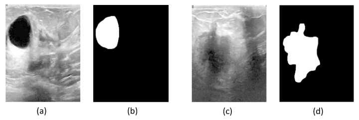
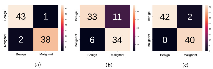
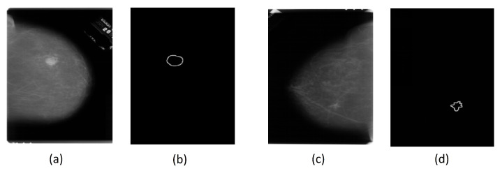
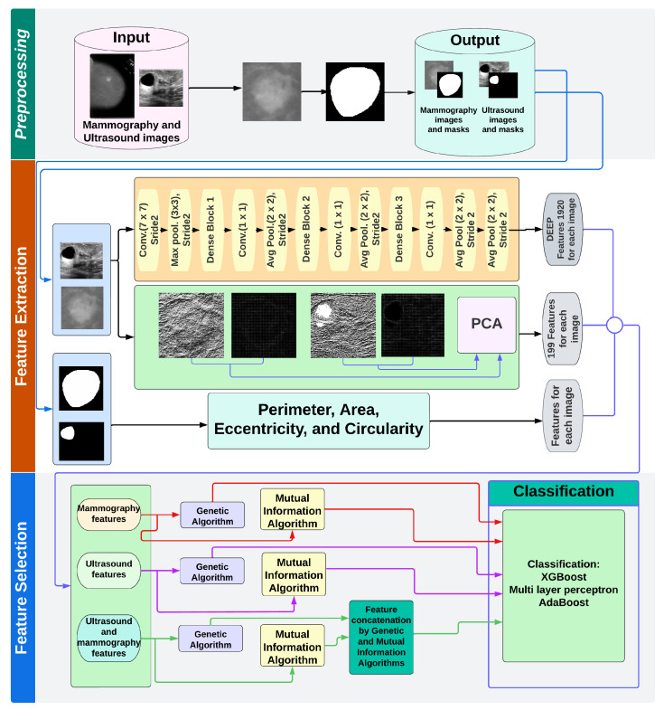
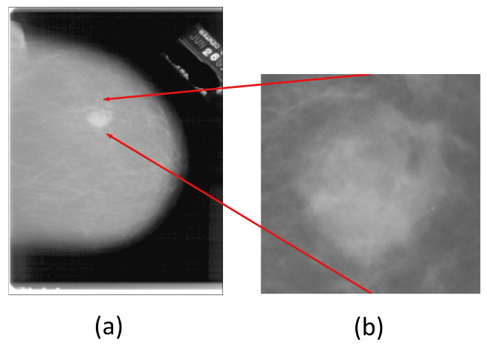
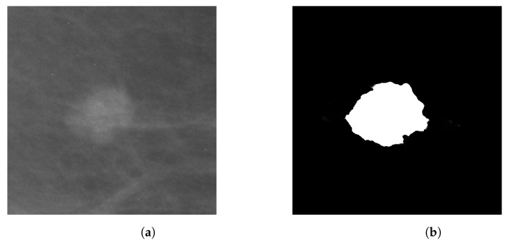
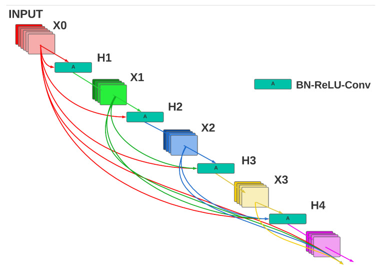
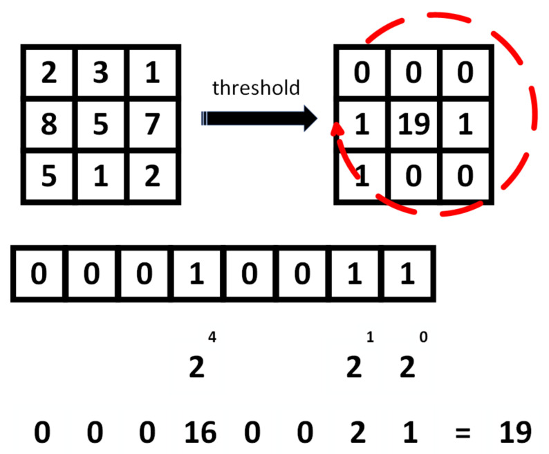
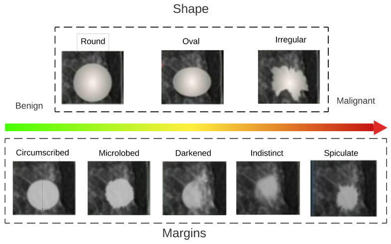
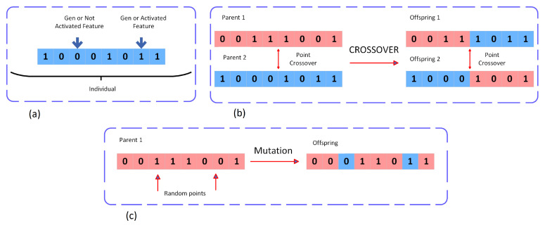
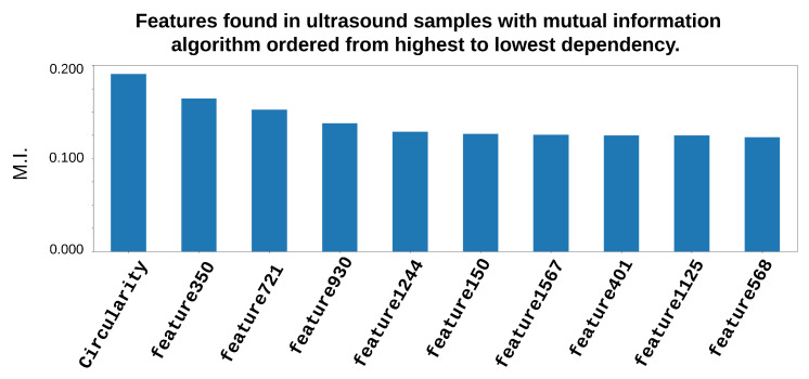
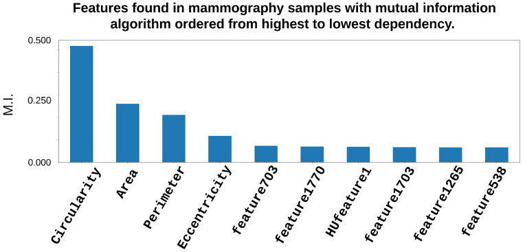
Breast cancer is a disease that affects women in different countries around the world. The real cause of breast cancer is particularly challenging to determine, and early detection of the disease is necessary for reducing the death rate, due to the high risks associated with breast cancer. Treatment in the early period can increase the life expectancy and quality of life for women. CAD (Computer Aided Diagnostic) systems can perform the diagnosis of the benign and malignant lesions of breast cancer using technologies and tools based on image processing, helping specialist doctors to obtain a more precise point of view with fewer processes when making their diagnosis by giving a second opinion. This study presents a novel CAD system for automated breast cancer diagnosis. The proposed method consists of different stages. In the preprocessing stage, an image is segmented, and a mask of a lesion is obtained; during the next stage, the extraction of the deep learning features is performed by a CNN-specifically, DenseNet 201. Additionally, handcrafted features (Histogram of Oriented Gradients (HOG)-based, ULBP-based, perimeter area, area, eccentricity, and circularity) are obtained from an image. The designed hybrid system uses CNN architecture for extracting deep learning features, along with traditional methods which perform several handcraft features, following the medical properties of the disease with the purpose of later fusion via proposed statistical criteria. During the fusion stage, where deep learning and handcrafted features are analyzed, the genetic algorithms as well as mutual information selection algorithm, followed by several classifiers (XGBoost, AdaBoost, Multilayer perceptron (MLP)) based on stochastic measures, are applied to choose the most sensible information group among the features. In the experimental validation of two modalities of the CAD design, which performed two types of medical studies-mammography (MG) and ultrasound (US)-the databases mini-DDSM (Digital Database for Screening Mammography) and BUSI (Breast Ultrasound Images Dataset) were used. Novel CAD systems were evaluated and compared with recent state-of-the-art systems, demonstrating better performance in commonly used criteria, obtaining ACC of 97.6%, PRE of 98%, Recall of 98%, F1-Score of 98%, and IBA of 95% for the abovementioned datasets.
乳腺癌是一种影响全球不同国家女性的疾病。确定乳腺癌的真正病因极具挑战性,由于乳腺癌相关风险高,早期发现该疾病对于降低死亡率至关重要。早期治疗可以提高女性的预期寿命和生活质量。计算机辅助诊断(CAD)系统可以使用基于图像处理的技术和工具对乳腺癌的良性和恶性病变进行诊断,通过提供第二种观点,帮助专科医生在进行诊断时以更少的流程获得更精确的观点。本研究提出了一种用于自动乳腺癌诊断的新型CAD系统。所提出的方法包括不同阶段。在预处理阶段,对图像进行分割,获得病变的掩码;在下一阶段,通过特定的卷积神经网络(CNN)——具体为DenseNet 201来提取深度学习特征。此外,从图像中获取手工特征(基于方向梯度直方图(HOG)、基于局部二值模式(ULBP)、周长面积、面积、偏心率和圆形度)。所设计的混合系统使用CNN架构来提取深度学习特征,同时结合传统方法来执行多个手工特征,遵循疾病的医学特性,以便稍后通过所提出的统计标准进行融合。在融合阶段,对深度学习和手工特征进行分析,应用遗传算法以及互信息选择算法,随后基于随机度量应用几个分类器(极端梯度提升(XGBoost)、自适应增强(AdaBoost)、多层感知器(MLP)),以在特征中选择最敏感的信息组。在对CAD设计的两种模态进行实验验证时,该设计进行了两种类型的医学研究——乳腺X线摄影(MG)和超声(US),使用了数据库小型数字乳腺X线摄影筛查数据库(mini-DDSM)和乳腺超声图像数据集(BUSI)。对新型CAD系统进行了评估,并与最近的先进系统进行了比较,在常用标准方面表现出更好的性能,对于上述数据集,准确率(ACC)为97.6%,精确率(PRE)为98%,召回率(Recall)为98%,F1分数为98%,综合平衡准确率(IBA)为95%。