• 文献检索
  • 文档翻译
  • 深度研究
  • 学术资讯
  • Suppr Zotero 插件Zotero 插件
  • 邀请有礼
  • 套餐&价格
  • 历史记录
应用&插件
Suppr Zotero 插件Zotero 插件浏览器插件Mac 客户端Windows 客户端微信小程序
定价
高级版会员购买积分包购买API积分包
服务
文献检索文档翻译深度研究API 文档MCP 服务
关于我们
关于 Suppr公司介绍联系我们用户协议隐私条款
关注我们

Suppr 超能文献

核心技术专利:CN118964589B侵权必究
粤ICP备2023148730 号-1Suppr @ 2026

文献检索

告别复杂PubMed语法,用中文像聊天一样搜索,搜遍4000万医学文献。AI智能推荐,让科研检索更轻松。

立即免费搜索

文件翻译

保留排版,准确专业,支持PDF/Word/PPT等文件格式,支持 12+语言互译。

免费翻译文档

深度研究

AI帮你快速写综述,25分钟生成高质量综述,智能提取关键信息,辅助科研写作。

立即免费体验

人工智能在乳腺癌检测中对乳腺病变的凸显作用。

Saliency of breast lesions in breast cancer detection using artificial intelligence.

机构信息

Escuela de Ingenierías Eléctrica Electrónica y de Telecomunicaciones, Universidad Industrial de Santander, Bucaramanga, Colombia.

Faculty of Medicine and Health Technology, Tampere University, Tampere, Finland.

出版信息

Sci Rep. 2023 Nov 23;13(1):20545. doi: 10.1038/s41598-023-46921-3.

DOI:10.1038/s41598-023-46921-3
PMID:37996504
原文链接:https://pmc.ncbi.nlm.nih.gov/articles/PMC10667547/
Abstract

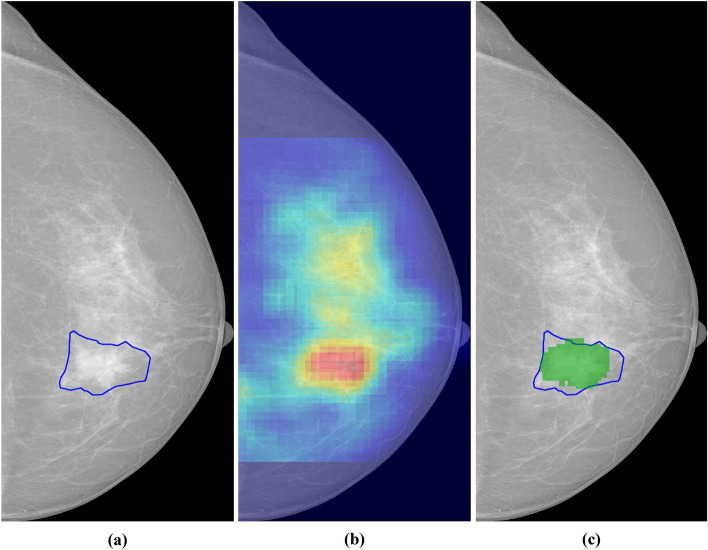
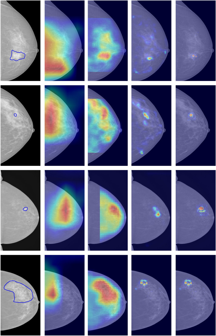
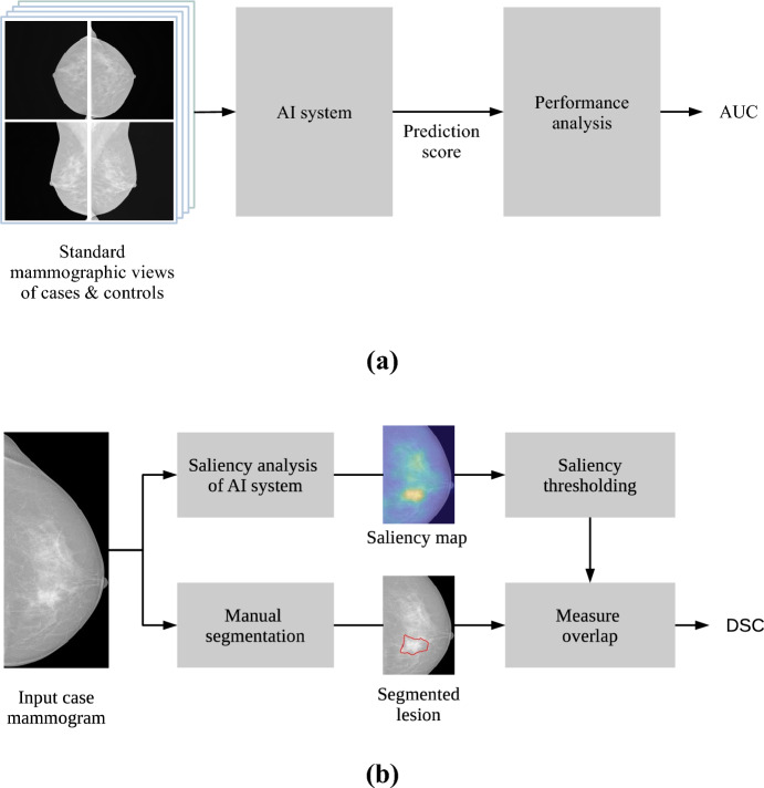
The analysis of mammograms using artificial intelligence (AI) has shown great potential for assisting breast cancer screening. We use saliency maps to study the role of breast lesions in the decision-making process of AI systems for breast cancer detection in screening mammograms. We retrospectively collected mammograms from 191 women with screen-detected breast cancer and 191 healthy controls matched by age and mammographic system. Two radiologists manually segmented the breast lesions in the mammograms from CC and MLO views. We estimated the detection performance of four deep learning-based AI systems using the area under the ROC curve (AUC) with a 95% confidence interval (CI). We used automatic thresholding on saliency maps from the AI systems to identify the areas of interest on the mammograms. Finally, we measured the overlap between these areas of interest and the segmented breast lesions using Dice's similarity coefficient (DSC). The detection performance of the AI systems ranged from low to moderate (AUCs from 0.525 to 0.694). The overlap between the areas of interest and the breast lesions was low for all the studied methods (median DSC from 4.2% to 38.0%). The AI system with the highest cancer detection performance (AUC = 0.694, CI 0.662-0.726) showed the lowest overlap (DSC = 4.2%) with breast lesions. The areas of interest found by saliency analysis of the AI systems showed poor overlap with breast lesions. These results suggest that AI systems with the highest performance do not solely rely on localized breast lesions for their decision-making in cancer detection; rather, they incorporate information from large image regions. This work contributes to the understanding of the role of breast lesions in cancer detection using AI.

摘要

使用人工智能(AI)分析乳房 X 光片在乳腺癌筛查中显示出很大的辅助作用。我们使用显著图来研究在用于筛查性乳房 X 光片中乳腺癌检测的 AI 系统的决策过程中,乳房病变的作用。我们回顾性地收集了 191 名经筛检发现患有乳腺癌的女性和 191 名年龄和乳房 X 光系统匹配的健康对照者的乳房 X 光片。两位放射科医生分别手动在 CC 和 MLO 视图的乳房 X 光片中对乳房病变进行了分割。我们使用接受者操作特征曲线(ROC)下面积(AUC)及其 95%置信区间(CI)来估计四个基于深度学习的 AI 系统的检测性能。我们使用 AI 系统的显著图自动阈值确定乳房 X 光片上的感兴趣区域。最后,我们使用 Dice 相似系数(DSC)测量这些感兴趣区域与分割的乳房病变之间的重叠。AI 系统的检测性能从低到中不等(AUC 从 0.525 到 0.694)。对于所有研究的方法,感兴趣区域与乳房病变之间的重叠都很低(中位数 DSC 从 4.2%到 38.0%)。癌症检测性能最高的 AI 系统(AUC=0.694,CI 0.662-0.726)与乳房病变的重叠最低(DSC=4.2%)。AI 系统的显著分析找到的感兴趣区域与乳房病变的重叠度很差。这些结果表明,性能最高的 AI 系统的决策不仅仅依赖于局部的乳房病变来进行癌症检测;相反,它们还整合了来自大图像区域的信息。这项工作有助于理解 AI 用于癌症检测中乳房病变的作用。

https://cdn.ncbi.nlm.nih.gov/pmc/blobs/ab36/10667547/c7bc4d6929ad/41598_2023_46921_Fig3_HTML.jpg
https://cdn.ncbi.nlm.nih.gov/pmc/blobs/ab36/10667547/8a7199a6e65e/41598_2023_46921_Fig1_HTML.jpg
https://cdn.ncbi.nlm.nih.gov/pmc/blobs/ab36/10667547/90e3012bc537/41598_2023_46921_Fig2_HTML.jpg
https://cdn.ncbi.nlm.nih.gov/pmc/blobs/ab36/10667547/c7bc4d6929ad/41598_2023_46921_Fig3_HTML.jpg
https://cdn.ncbi.nlm.nih.gov/pmc/blobs/ab36/10667547/8a7199a6e65e/41598_2023_46921_Fig1_HTML.jpg
https://cdn.ncbi.nlm.nih.gov/pmc/blobs/ab36/10667547/90e3012bc537/41598_2023_46921_Fig2_HTML.jpg
https://cdn.ncbi.nlm.nih.gov/pmc/blobs/ab36/10667547/c7bc4d6929ad/41598_2023_46921_Fig3_HTML.jpg

相似文献

1
Saliency of breast lesions in breast cancer detection using artificial intelligence.人工智能在乳腺癌检测中对乳腺病变的凸显作用。
Sci Rep. 2023 Nov 23;13(1):20545. doi: 10.1038/s41598-023-46921-3.
2
Screening mammography performance according to breast density: a comparison between radiologists versus standalone intelligence detection.根据乳腺密度评估乳腺钼靶筛查性能:放射科医生与独立智能检测的比较。
Breast Cancer Res. 2024 Apr 22;26(1):68. doi: 10.1186/s13058-024-01821-w.
3
AI for interpreting screening mammograms: implications for missed cancer in double reading practices and challenging-to-locate lesions.人工智能解读乳腺 X 光筛查片:对双读实践中漏诊癌症和难以定位病变的影响。
Sci Rep. 2024 May 24;14(1):11893. doi: 10.1038/s41598-024-62324-4.
4
Performance of an Artificial Intelligence System for Breast Cancer Detection on Screening Mammograms from BreastScreen Norway.基于挪威乳腺筛查项目的筛查性乳腺钼靶 X 光片评估乳腺癌检测人工智能系统的性能。
Radiol Artif Intell. 2024 May;6(3):e230375. doi: 10.1148/ryai.230375.
5
An Artificial Intelligence-based Mammography Screening Protocol for Breast Cancer: Outcome and Radiologist Workload.基于人工智能的乳腺癌 X 光筛查协议:结果和放射科医生的工作量。
Radiology. 2022 Jul;304(1):41-49. doi: 10.1148/radiol.210948. Epub 2022 Apr 19.
6
Accuracy of an Artificial Intelligence System for Interval Breast Cancer Detection at Screening Mammography.人工智能系统在筛查性乳房 X 光摄影中检测间期乳腺癌的准确性。
Radiology. 2024 Aug;312(2):e232303. doi: 10.1148/radiol.232303.
7
Artificial intelligence in BreastScreen Norway: a retrospective analysis of a cancer-enriched sample including 1254 breast cancer cases.挪威乳腺 X 线筛查中的人工智能:包括 1254 例乳腺癌病例的癌症富集样本的回顾性分析。
Eur Radiol. 2023 May;33(5):3735-3743. doi: 10.1007/s00330-023-09461-y. Epub 2023 Mar 14.
8
How do AI markings on screening mammograms correspond to cancer location? An informed review of 270 breast cancer cases in BreastScreen Norway.乳腺 X 线筛查片上的人工智能标记与癌症位置如何对应?对 270 例挪威乳腺筛查乳腺癌病例的知情审查。
Eur Radiol. 2024 Sep;34(9):6158-6167. doi: 10.1007/s00330-024-10662-2. Epub 2024 Feb 23.
9
Artificial intelligence (AI) for breast cancer screening: BreastScreen population-based cohort study of cancer detection.人工智能(AI)在乳腺癌筛查中的应用:基于乳腺筛查人群队列的癌症检测研究。
EBioMedicine. 2023 Apr;90:104498. doi: 10.1016/j.ebiom.2023.104498. Epub 2023 Feb 28.
10
Diagnostic Performance of AI for Cancers Registered in A Mammography Screening Program: A Retrospective Analysis.人工智能在乳腺筛查项目中用于癌症诊断的性能:一项回顾性分析。
Technol Cancer Res Treat. 2022 Jan-Dec;21:15330338221075172. doi: 10.1177/15330338221075172.

引用本文的文献

1
Optimizing Artificial Intelligence Thresholds for Mammographic Lesion Detection: A Retrospective Study on Diagnostic Performance and Radiologist-Artificial Intelligence Discordance.优化用于乳腺钼靶病变检测的人工智能阈值:一项关于诊断性能及放射科医生与人工智能诊断不一致性的回顾性研究
Diagnostics (Basel). 2025 May 29;15(11):1368. doi: 10.3390/diagnostics15111368.
2
The utility of quantifying the orientation of breast masses in ultrasound imaging.定量超声成像中乳房肿块方位的应用。
Sci Rep. 2024 Feb 25;14(1):4578. doi: 10.1038/s41598-024-55298-w.

本文引用的文献

1
Artificial Intelligence as Supporting Reader in Breast Screening: A Novel Workflow to Preserve Quality and Reduce Workload.人工智能作为乳腺筛查的辅助阅读工具:一种既能保证质量又能减轻工作量的新工作流程。
J Breast Imaging. 2023 May 22;5(3):267-276. doi: 10.1093/jbi/wbad010.
2
Impact of Different Mammography Systems on Artificial Intelligence Performance in Breast Cancer Screening.不同乳腺钼靶摄影系统对乳腺癌筛查中人工智能性能的影响。
Radiol Artif Intell. 2023 Mar 22;5(3):e220146. doi: 10.1148/ryai.220146. eCollection 2023 May.
3
Standalone AI for Breast Cancer Detection at Screening Digital Mammography and Digital Breast Tomosynthesis: A Systematic Review and Meta-Analysis.
筛查数字乳腺 X 线摄影和数字乳腺断层合成术中用于乳腺癌检测的独立人工智能:系统评价和荟萃分析。
Radiology. 2023 Jun;307(5):e222639. doi: 10.1148/radiol.222639. Epub 2023 May 23.
4
ADMANI: Annotated Digital Mammograms and Associated Non-Image Datasets.ADMANI:带注释的数字乳腺X线照片及相关非图像数据集。
Radiol Artif Intell. 2022 Dec 21;5(2):e220072. doi: 10.1148/ryai.220072. eCollection 2023 Mar.
5
Explainable AI in medical imaging: An overview for clinical practitioners - Saliency-based XAI approaches.可解释人工智能在医学影像中的应用:临床医师的概述——基于显著度的 XAI 方法。
Eur J Radiol. 2023 May;162:110787. doi: 10.1016/j.ejrad.2023.110787. Epub 2023 Mar 21.
6
An in silico study on the detectability of field cancerization through parenchymal analysis of digital mammograms.数字乳腺钼靶图像实质分析对癌前病变检测能力的计算机模拟研究
Med Phys. 2023 Oct;50(10):6379-6389. doi: 10.1002/mp.16401. Epub 2023 Apr 21.
7
The EMory BrEast imaging Dataset (EMBED): A Racially Diverse, Granular Dataset of 3.4 Million Screening and Diagnostic Mammographic Images.埃默里乳腺成像数据集(EMBED):一个包含340万张筛查和诊断性乳腺钼靶图像的种族多样化、详细的数据集。
Radiol Artif Intell. 2023 Jan 4;5(1):e220047. doi: 10.1148/ryai.220047. eCollection 2023 Jan.
8
A systematic review on the use of explainability in deep learning systems for computer aided diagnosis in radiology: Limited use of explainable AI?关于深度学习系统在放射学计算机辅助诊断中应用的可解释性的系统评价:可解释人工智能的使用有限?
Eur J Radiol. 2022 Dec;157:110592. doi: 10.1016/j.ejrad.2022.110592. Epub 2022 Nov 5.
9
Towards a guideline for evaluation metrics in medical image segmentation.迈向医学图像分割评估指标指南。
BMC Res Notes. 2022 Jun 20;15(1):210. doi: 10.1186/s13104-022-06096-y.
10
Explainable artificial intelligence (XAI) in deep learning-based medical image analysis.深度学习在医学影像分析中的可解释人工智能(XAI)。
Med Image Anal. 2022 Jul;79:102470. doi: 10.1016/j.media.2022.102470. Epub 2022 May 4.