Department of Surgery, Stomatology, Pathology and Radiology, Bauru School of Dentistry. University of São Paulo, Alameda Dr. Octávio Pinheiro Brisolla, 9-75, Bauru, SP, 17012-901, Brazil.
BMC Oral Health. 2023 Nov 23;23(1):915. doi: 10.1186/s12903-023-03653-0.
Lower third molars (L3M) are the last teeth to erupt in the oral cavity. Uneruption of these teeth still raises questions about its causes, in the literature (1) genetic factors, (2) dental lamina activity and, mainly, (3) insufficient growth and development of the bone bases are included. While the lack of space theory influenced by mandibular morphology and size of L3M was argued to be the main reason for L3M impaction, there is a limitation in the literature in examining such association using more accurate tomographic analysis obtained from CBCT. This work aimed to evaluate the relationship between mandibular morphology and the eruption of L3M.
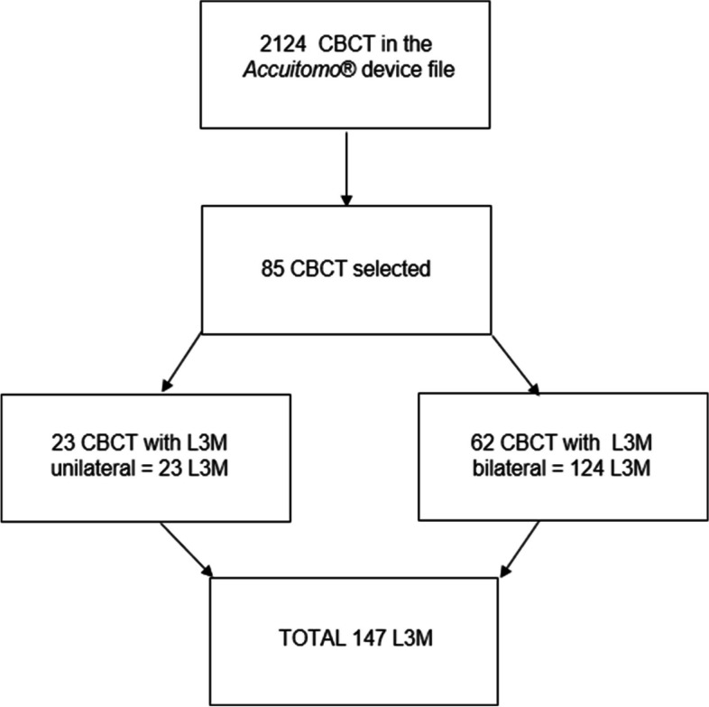
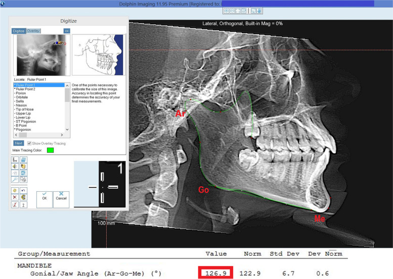
In this regard, 85 Cone Beam Computed Tomographies (CBCT), with 147 L3M, were selected from the archives of the Department of Surgery, Stomatology, Pathology and Radiology, Bauru School of Dentistry, obtained using an Accuitomo® Morita device and using the Dolphin Imaging 11.9 software. L3M eruption was related to linear measurements of jaw length (Co-Gn), retromolar space dimension (D2R), mesiodistal width of the L3M crowns, mandibular first molars (L1M) and mandibular canines (LC) and the angle mandibular (Ar-Go-Me). Independent samples t-test, chi-square tests and logistic regression were performed adopting a significance level of 5%.
The average mandible length of 116.446 mm + 6.415 mm, retromolar space of 11.634 mm + 2.385 mm, mesiodistal size of the L3M of 10.054 mm + 0.941 mm, sum of the mesiodistal widths of the L1M and LC of 15.564 mm + 1.218 mm and mandibular angle of 127.23° + 6.109. There was no statistically significant association between these factors and the eruption.
With the results obtained in this study, we conclude that the length and angle of the mandible, teeth size and dimension of the retromolar space are not associated with the L3M eruption.
下颌第三磨牙(L3M)是口腔中最后萌出的牙齿。这些牙齿的萌出情况仍然存在争议,文献中提到了(1)遗传因素、(2)牙板活动以及(3)骨基底生长发育不足等原因。尽管下颌形态和 L3M 大小受下颌缺乏空间理论的影响被认为是 L3M 阻生的主要原因,但文献中使用更准确的 CBCT 断层分析来检查这种关联存在局限性。本研究旨在评估下颌形态与 L3M 萌出的关系。
为此,从巴鲁牙科外科、口腔医学、病理学和放射学系的档案中选择了 85 例锥形束 CT(CBCT),共 147 颗 L3M,使用 Accuitomo® Morita 设备获取,并使用 Dolphin Imaging 11.9 软件。L3M 的萌出与下颌长度(Co-Gn)、磨牙后间隙(D2R)、L3M 牙冠的近远中宽度、下颌第一磨牙(L1M)和下颌尖牙(LC)以及下颌角(Ar-Go-Me)的线性测量值相关。采用独立样本 t 检验、卡方检验和逻辑回归分析,显著性水平为 5%。
平均下颌长度为 116.446 mm+6.415 mm,磨牙后间隙为 11.634 mm+2.385 mm,L3M 的近远中尺寸为 10.054 mm+0.941 mm,L1M 和 LC 的近远中宽度之和为 15.564 mm+1.218 mm,下颌角为 127.23°+6.109°。这些因素与萌出之间无统计学显著相关性。
根据本研究的结果,我们得出结论,下颌的长度和角度、牙齿大小以及磨牙后间隙的尺寸与 L3M 的萌出无关。