• 文献检索
  • 文档翻译
  • 深度研究
  • 学术资讯
  • Suppr Zotero 插件Zotero 插件
  • 邀请有礼
  • 套餐&价格
  • 历史记录
应用&插件
Suppr Zotero 插件Zotero 插件浏览器插件Mac 客户端Windows 客户端微信小程序
定价
高级版会员购买积分包购买API积分包
服务
文献检索文档翻译深度研究API 文档MCP 服务
关于我们
关于 Suppr公司介绍联系我们用户协议隐私条款
关注我们

Suppr 超能文献

核心技术专利:CN118964589B侵权必究
粤ICP备2023148730 号-1Suppr @ 2026

文献检索

告别复杂PubMed语法,用中文像聊天一样搜索,搜遍4000万医学文献。AI智能推荐,让科研检索更轻松。

立即免费搜索

文件翻译

保留排版,准确专业,支持PDF/Word/PPT等文件格式,支持 12+语言互译。

免费翻译文档

深度研究

AI帮你快速写综述,25分钟生成高质量综述,智能提取关键信息,辅助科研写作。

立即免费体验

递送微小RNA-302a-3p的3D打印TCP-HA支架可改善小鼠颅骨模型中的骨再生。

3D-printed TCP-HA scaffolds delivering MicroRNA-302a-3p improve bone regeneration in a mouse calvarial model.

作者信息

Limlawan Pirawish, Insin Numpon, Marger Laurine, Freudenreich Mélanie, Durual Stéphane, Vacharaksa Anjalee

机构信息

Department of Oral Medicine, Faculty of Dentistry, Chulalongkorn University, Pathumwan, Bangkok, 10330, Thailand.

Research Unit on Oral Microbiology and Immunology, Faculty of Dentistry, Chulalongkorn University, Pathumwan, Bangkok, 10330, Thailand.

出版信息

BDJ Open. 2023 Nov 24;9(1):50. doi: 10.1038/s41405-023-00177-1.

DOI:10.1038/s41405-023-00177-1
PMID:38001073
原文链接:https://pmc.ncbi.nlm.nih.gov/articles/PMC10673873/
Abstract

OBJECTIVE

To demonstrate hydroxyapatite nanoparticles modified with cationic functional molecules. 3-aminopropyltriethoxysilane (HA-NPs-APTES) carrying microRNA-302a-3p (miR) in the 3D-printed tricalcium phosphate/Hydroxyapatite (TCP/HA) scaffold can increase healing of the critical-sized bone defect.

MATERIALS AND METHODS

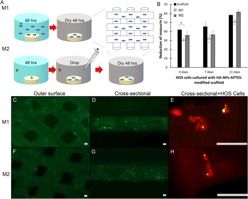
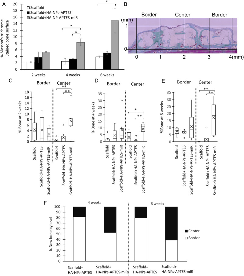
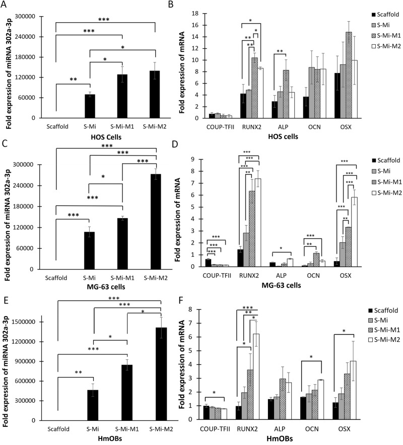
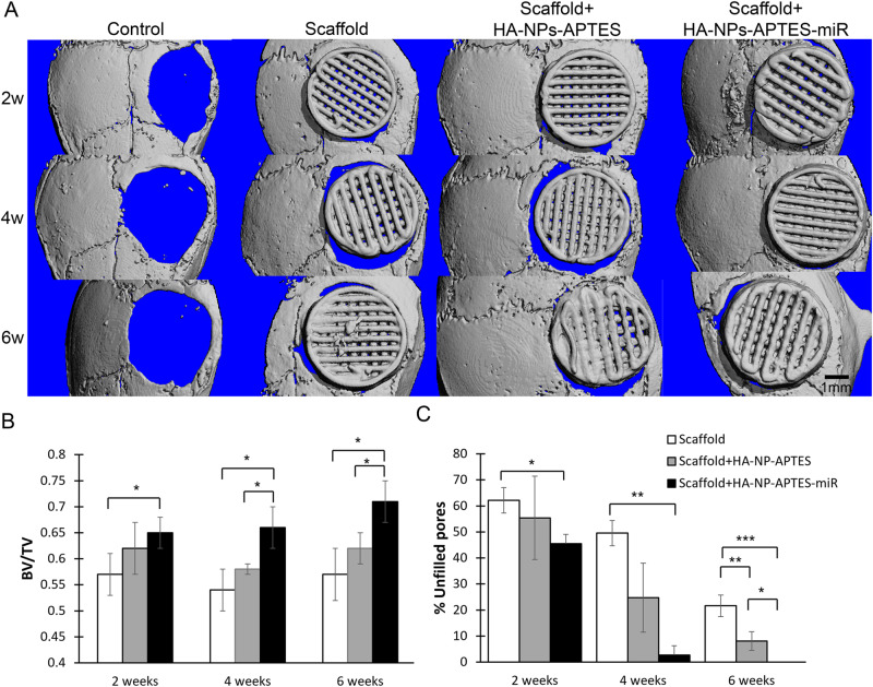
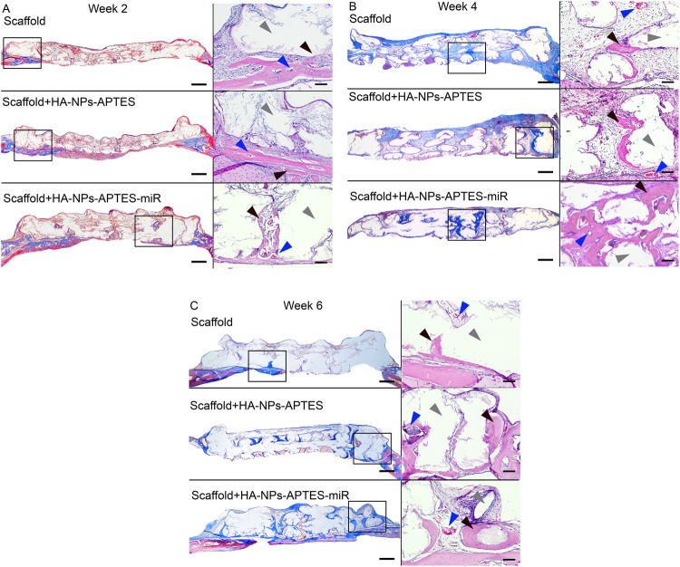
3D-printed TCP/HA were modified with HA-NPs-APTES by two methods (M1, M2). The dispersion of particles was visualized by fluorescent microscopy. Biocompatibility of the scaffolds was tested by alizarin assay. Delivery of miR to the cells and osteogenic gene expression were evaluated by qPCR. After selecting best method (M2), scaffolds, scaffolds+HA-NPs-APTES with or without miR were implanted in 4 mm mouse calvarium defect (n = 4 per group). After 2,4 and 6 weeks, bone regeneration were evaluated by microCT and histology sections.

RESULTS

Both M1 and M2 scaffolds were biocompatible with cell adhesion on its surface. M2 scaffold showed significant increase of miR, suggesting successful delivery, resulted in downregulation of its target mRNA COUP-TFII, and upregulation of RUNX2 mRNA. Calvarium defect with M2 scaffold also showed significantly higher BV/TV and higher number of filled spaces at all time points. Histomorphometry demonstrated new bone formed at the center of the HA-NPs-APTES-miR scaffold earlier than controls.

CONCLUSION

TCP/HA scaffold modified with HA-NPs-APTES facilitated delivery of miR and enhanced bone regeneration.

摘要

目的

展示用阳离子功能分子修饰的羟基磷灰石纳米颗粒。在3D打印的磷酸三钙/羟基磷灰石(TCP/HA)支架中携带微小RNA-302a-3p(miR)的3-氨丙基三乙氧基硅烷(HA-NPs-APTES)可促进临界尺寸骨缺损的愈合。

材料与方法

采用两种方法(M1、M2)用HA-NPs-APTES对3D打印的TCP/HA进行修饰。通过荧光显微镜观察颗粒的分散情况。通过茜素试验测试支架的生物相容性。通过qPCR评估miR向细胞的递送和骨生成基因表达。在选择最佳方法(M2)后,将支架、含或不含miR的支架+HA-NPs-APTES植入4毫米的小鼠颅骨缺损处(每组n = 4)。在2、4和6周后,通过显微CT和组织学切片评估骨再生情况。

结果

M1和M2支架均具有生物相容性,细胞可在其表面黏附。M2支架显示miR显著增加,表明递送成功,导致其靶mRNA COUP-TFII下调,RUNX2 mRNA上调。植入M2支架后的颅骨缺损在所有时间点的骨体积分数(BV/TV)也显著更高,填充空间数量更多。组织形态计量学显示,HA-NPs-APTES-miR支架中心形成新骨的时间早于对照组。

结论

用HA-NPs-APTES修饰的TCP/HA支架促进了miR的递送并增强了骨再生。

https://cdn.ncbi.nlm.nih.gov/pmc/blobs/0143/10673873/98f33cc962bc/41405_2023_177_Fig5_HTML.jpg
https://cdn.ncbi.nlm.nih.gov/pmc/blobs/0143/10673873/422afcfafcb8/41405_2023_177_Fig1_HTML.jpg
https://cdn.ncbi.nlm.nih.gov/pmc/blobs/0143/10673873/af0e843a304c/41405_2023_177_Fig2_HTML.jpg
https://cdn.ncbi.nlm.nih.gov/pmc/blobs/0143/10673873/c46fd3597c8b/41405_2023_177_Fig3_HTML.jpg
https://cdn.ncbi.nlm.nih.gov/pmc/blobs/0143/10673873/7beb5cdc5094/41405_2023_177_Fig4_HTML.jpg
https://cdn.ncbi.nlm.nih.gov/pmc/blobs/0143/10673873/98f33cc962bc/41405_2023_177_Fig5_HTML.jpg

相似文献

1
3D-printed TCP-HA scaffolds delivering MicroRNA-302a-3p improve bone regeneration in a mouse calvarial model.递送微小RNA-302a-3p的3D打印TCP-HA支架可改善小鼠颅骨模型中的骨再生。
BDJ Open. 2023 Nov 24;9(1):50. doi: 10.1038/s41405-023-00177-1.
2
Delivery of microRNA-302a-3p by APTES modified hydroxyapatite nanoparticles to promote osteogenic differentiation in vitro.通过APTES修饰的羟基磷灰石纳米颗粒递送微小RNA-302a-3p以促进体外成骨分化。
BDJ Open. 2023 Feb 22;9(1):8. doi: 10.1038/s41405-023-00135-x.
3
Bone regeneration in critical bone defects using three-dimensionally printed β-tricalcium phosphate/hydroxyapatite scaffolds is enhanced by coating scaffolds with either dipyridamole or BMP-2.使用三维打印的β-磷酸三钙/羟基磷灰石支架修复严重骨缺损时,通过用双嘧达莫或骨形态发生蛋白-2包被支架可增强骨再生。
J Biomed Mater Res B Appl Biomater. 2017 Feb;105(2):366-375. doi: 10.1002/jbm.b.33561. Epub 2015 Oct 29.
4
Early In Vivo Osteogenic and Inflammatory Response of 3D Printed Polycaprolactone/Carbon Nanotube/Hydroxyapatite/Tricalcium Phosphate Composite Scaffolds.3D打印聚己内酯/碳纳米管/羟基磷灰石/磷酸三钙复合支架的早期体内成骨和炎症反应
Polymers (Basel). 2023 Jul 5;15(13):2952. doi: 10.3390/polym15132952.
5
Rat Calvarial Bone Regeneration by 3D-Printed β-Tricalcium Phosphate Incorporating MicroRNA-200c.3D 打印β-磷酸三钙载 miRNA-200c 促进大鼠颅骨再生
ACS Biomater Sci Eng. 2021 Sep 13;7(9):4521-4534. doi: 10.1021/acsbiomaterials.0c01756. Epub 2021 Aug 26.
6
Chitosan/gelatin/platelet gel enriched by a combination of hydroxyapatite and beta-tricalcium phosphate in healing of a radial bone defect model in rat.壳聚糖/明胶/血小板凝胶通过羟基磷灰石和β-磷酸三钙的组合富集用于大鼠桡骨缺损模型的愈合
Int J Biol Macromol. 2017 Aug;101:630-637. doi: 10.1016/j.ijbiomac.2017.03.148. Epub 2017 Mar 28.
7
The stimulation of healing within a rat calvarial defect by mPCL-TCP/collagen scaffolds loaded with rhBMP-2.负载重组人骨形态发生蛋白-2的微孔隙聚己内酯-磷酸三钙/胶原蛋白支架对大鼠颅骨缺损愈合的刺激作用
Biomaterials. 2009 May;30(13):2479-88. doi: 10.1016/j.biomaterials.2008.12.055. Epub 2009 Jan 21.
8
Three dimensional printed macroporous polylactic acid/hydroxyapatite composite scaffolds for promoting bone formation in a critical-size rat calvarial defect model.用于促进大鼠颅骨临界尺寸缺损模型骨形成的三维打印大孔聚乳酸/羟基磷灰石复合支架
Sci Technol Adv Mater. 2016 Apr 8;17(1):136-148. doi: 10.1080/14686996.2016.1145532. eCollection 2016.
9
VEGF-Loaded Heparinised Gelatine-Hydroxyapatite-Tricalcium Phosphate Scaffold Accelerates Bone Regeneration Enhancing Osteogenesis-Angiogenesis Coupling.负载血管内皮生长因子的肝素化明胶-羟基磷灰石-磷酸三钙支架通过增强成骨-血管生成耦合加速骨再生。
Front Bioeng Biotechnol. 2022 Jun 8;10:915181. doi: 10.3389/fbioe.2022.915181. eCollection 2022.
10
Regional gene therapy with 3D printed scaffolds to heal critical sized bone defects in a rat model.利用 3D 打印支架进行区域性基因治疗,以治愈大鼠模型中的临界尺寸骨缺损。
J Biomed Mater Res A. 2019 Oct;107(10):2174-2182. doi: 10.1002/jbm.a.36727. Epub 2019 Jun 11.

引用本文的文献

1
MicroRNA functions in osteogenic differentiation of periodontal ligament stem cells: a scoping review.微小RNA在牙周膜干细胞成骨分化中的作用:一项范围综述
Front Oral Health. 2025 Jan 31;6:1423226. doi: 10.3389/froh.2025.1423226. eCollection 2025.
2
MicroRNA-21-5p profile in the alveolar bone following tooth extraction in medication-related osteonecrosis of the jaw rat model.药物相关性颌骨坏死大鼠模型拔牙后牙槽骨中的微小RNA-21-5p表达情况
Front Dent Med. 2024 Dec 17;5:1477274. doi: 10.3389/fdmed.2024.1477274. eCollection 2024.
3
Current advances in the development of microRNA-integrated tissue engineering strategies: a cornerstone of regenerative medicine.

本文引用的文献

1
Alveolar ridge augmentation with 3D-printed synthetic bone blocks: A clinical case series.使用3D打印合成骨块进行牙槽嵴增高术:临床病例系列
Clin Case Rep. 2023 Apr 23;11(4):e7171. doi: 10.1002/ccr3.7171. eCollection 2023 Apr.
2
Delivery of microRNA-302a-3p by APTES modified hydroxyapatite nanoparticles to promote osteogenic differentiation in vitro.通过APTES修饰的羟基磷灰石纳米颗粒递送微小RNA-302a-3p以促进体外成骨分化。
BDJ Open. 2023 Feb 22;9(1):8. doi: 10.1038/s41405-023-00135-x.
3
Systemic proteomics and miRNA profile analysis of exosomes derived from human pluripotent stem cells.
微小RNA整合组织工程策略发展的当前进展:再生医学的基石
Front Bioeng Biotechnol. 2024 Oct 16;12:1484151. doi: 10.3389/fbioe.2024.1484151. eCollection 2024.
人多能干细胞来源的外泌体的系统蛋白质组学和 miRNA 谱分析。
Stem Cell Res Ther. 2022 Sep 5;13(1):449. doi: 10.1186/s13287-022-03142-1.
4
VEGFA from osteoblasts is not required for lamellar bone formation following tibial loading.成骨细胞中的 VEGFA 对于胫骨加载后的板层骨形成不是必需的。
Bone. 2022 Oct;163:116502. doi: 10.1016/j.bone.2022.116502. Epub 2022 Jul 21.
5
Oxidative-Induced Angiogenesis Is Modulated by Small Extracellular Vesicle miR-302a-3p Cargo in Retinal Pigment Epithelium Cells.氧化诱导的血管生成受视网膜色素上皮细胞中小细胞外囊泡miR-302a-3p货物的调节。
Antioxidants (Basel). 2022 Apr 22;11(5):818. doi: 10.3390/antiox11050818.
6
Exosomal microRNA‑302a promotes trophoblast migration and proliferation, and represses angiogenesis by regulating the expression levels of VEGFA in preeclampsia.外泌体 microRNA-302a 通过调节子痫前期中 VEGFA 的表达水平,促进滋养细胞迁移和增殖,并抑制血管生成。
Mol Med Rep. 2021 Dec;24(6). doi: 10.3892/mmr.2021.12504. Epub 2021 Oct 22.
7
Rat Calvarial Bone Regeneration by 3D-Printed β-Tricalcium Phosphate Incorporating MicroRNA-200c.3D 打印β-磷酸三钙载 miRNA-200c 促进大鼠颅骨再生
ACS Biomater Sci Eng. 2021 Sep 13;7(9):4521-4534. doi: 10.1021/acsbiomaterials.0c01756. Epub 2021 Aug 26.
8
MiR-302a Limits Vascular Inflammation by Suppressing Nuclear Factor-κ B Pathway in Endothelial Cells.微小RNA-302a通过抑制内皮细胞中的核因子-κB通路来限制血管炎症。
Front Cell Dev Biol. 2021 Aug 2;9:682574. doi: 10.3389/fcell.2021.682574. eCollection 2021.
9
MicroRNA-302a-3p induces ferroptosis of non-small cell lung cancer cells via targeting ferroportin.微小RNA-302a-3p通过靶向铁转运蛋白诱导非小细胞肺癌细胞发生铁死亡。
Free Radic Res. 2021 Jul;55(7):821-830. doi: 10.1080/10715762.2021.1947503. Epub 2021 Aug 6.
10
Advancing application of mesenchymal stem cell-based bone tissue regeneration.基于间充质干细胞的骨组织再生的应用进展。
Bioact Mater. 2020 Sep 21;6(3):666-683. doi: 10.1016/j.bioactmat.2020.08.014. eCollection 2021 Mar.