State Key Laboratory of Ophthalmology, Zhongshan Ophthalmic Center, Sun Yat-Sen University, Guangdong Provincial Key Laboratory of Ophthalmology and Visual Science, Guangzhou, China.
Yale College, Yale University, New Haven, Connecticut, United States.
Invest Ophthalmol Vis Sci. 2023 Nov 1;64(14):42. doi: 10.1167/iovs.64.14.42.
This study aimed to explore the impact of GSK840 on retinal neuronal injury after retinal ischemia/reperfusion (IR) and its associated mechanism.
We established an in vivo mouse model of IR and an in vitro model of oxygen and glucose deprivation/reoxygenation (OGDR) in primary mouse retinal ganglion cells (RGCs). GSK840, a small-molecule compound, was used to specifically inhibit RIPK3/MLKL-dependent necroptosis. Retinal structure and function evaluation was performed by using hematoxylin and eosin staining, optical coherence tomography, and electroretinography. Propidium Iodide (PI) staining was used for detection of necroptotic cell death, whereas Western blot analysis and immunofluorescence were used to assess necroptosis-related proteins and inner retinal neurons.
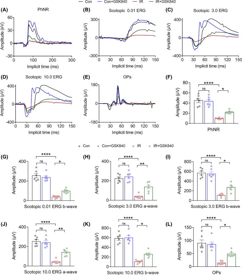
RIPK3/MLKL-dependent necroptosis was rapidly activated in RGCs following retinal IR or OGDR. GSK840 helped maintain relatively normal inner retinal structure and thickness by preserving inner retinal neurons, particularly RGCs. Meanwhile, GSK840 ameliorated IR-induced visual dysfunction, as evidenced by the improved amplitudes of photopic negative response, a-wave, b-wave, and oscillatory potentials. And GSK840 treatment significantly reduced the population of PI+ RGCs after injury. Mechanistically, GSK840 ameliorated RGC necroptosis by inhibiting the RIPK3/MLKL pathway.
GSK840 exerts protective effects against retinal neuronal injury after IR by inhibiting RIPK3/MLKL-mediated RGC necroptosis. GSK840 may represent a protective strategy for RGC degeneration in ischemic retinopathy.
本研究旨在探讨 GSK840 对视网膜缺血/再灌注(IR)后神经元损伤的影响及其相关机制。
我们建立了体内小鼠 IR 模型和原代小鼠视网膜神经节细胞(RGC)体外氧葡萄糖剥夺/再氧合(OGDR)模型。GSK840 是一种小分子化合物,可特异性抑制 RIPK3/MLKL 依赖性坏死性凋亡。采用苏木精和伊红染色、光相干断层扫描和视网膜电图评估视网膜结构和功能。碘化丙啶(PI)染色用于检测坏死性凋亡细胞死亡,而 Western blot 分析和免疫荧光用于评估坏死性凋亡相关蛋白和内层视网膜神经元。
在视网膜 IR 或 OGDR 后,RGC 中迅速激活 RIPK3/MLKL 依赖性坏死性凋亡。GSK840 通过维持内层视网膜神经元,特别是 RGC 的相对正常的内层视网膜结构和厚度,有助于维持相对正常的内层视网膜结构和厚度。同时,GSK840 改善了 IR 诱导的视觉功能障碍,表现为光峰负反应、a 波、b 波和振荡电位的振幅改善。并且 GSK840 处理后损伤后 PI+RGC 的数量显著减少。机制上,GSK840 通过抑制 RIPK3/MLKL 通路改善 RGC 坏死性凋亡。
GSK840 通过抑制 RIPK3/MLKL 介导的 RGC 坏死性凋亡,对 IR 后视网膜神经元损伤发挥保护作用。GSK840 可能代表缺血性视网膜病变中 RGC 变性的一种保护策略。