Centre for Optometry and Vision Science, Biomedical Sciences Research Institute, Ulster University, Coleraine, UK.
National Institute for Health Research Moorfields Biomedical Research Centre, Moorfields Eye Hospital NHS Foundation Trust and UCL Institute of Ophthalmology, London, UK.
Transl Vis Sci Technol. 2023 Nov 1;12(11):37. doi: 10.1167/tvst.12.11.37.
To measure achromatic spatial, temporal, and spatiotemporal summation in dry age-related macular degeneration (AMD) compared to healthy controls under conditions of photopic gaze-contingent perimetry.
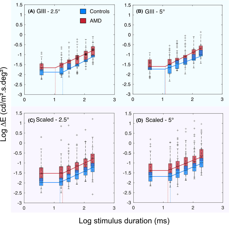
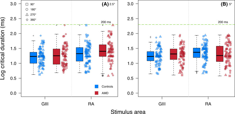
Twenty participants with dry AMD (mean age, 74.6 years) and 20 healthy controls (mean age, 67.8 years) performed custom, gaze-contingent perimetry tests. An area-modulation test generated localized estimates of Ricco's area (RA) at 2.5° and 5° eccentricities along the 0°, 90°, 180°, and 270° meridians. Contrast thresholds were measured at the same test locations for stimuli of six durations (3.7-190.4 ms) with a Goldmann III stimulus (GIII, 0.43°) and RA-scaled stimuli. The upper limit (critical duration) of complete temporal summation (using the GIII stimulus) and spatiotemporal summation (using the RA stimuli) was estimated using iterative two-phase regression analysis.
Median (interquartile range [IQR]) RA estimates were significantly larger in AMD participants (2.5°: 0.21 [0.09-0.41] deg2; 5°: 0.32 [0.15-0.65 deg2]) compared to healthy controls (2.5°: 0.08 [0.05-0.13] deg2; 5°: 0.15 [0.08-0.22] deg2) at all test locations (all P < 0.05). No significant difference in median critical duration was found in AMD participants with the GIII stimulus (19.6 [9.9-30.4] ms) and RA-scaled stimuli (22.9 [13.9-40.3] ms) compared to healthy controls (GIII: 17.0 [11.3-24.0] ms; RA-scaled: 22.4 [14.3-33.1] ms) at all test locations (all P > 0.05).
Spatial summation is altered in dry AMD, without commensurate changes in temporal summation.
The sensitivity of perimetry to AMD may be improved by utilizing stimuli that probe alterations in spatial summation in the disease.
在明视条件下,通过注视相关的光幻视周边视野检查,测量与健康对照相比,干性年龄相关性黄斑变性(AMD)患者的无彩空间、时间和时空总和。
20 名干性 AMD 患者(平均年龄 74.6 岁)和 20 名健康对照者(平均年龄 67.8 岁)进行了定制的、注视相关的周边视野检查。面积调制测试在 0°、90°、180°和 270°子午线上的 2.5°和 5°偏心处生成 Ricco 区域(RA)的局部估计值。使用 Goldmann III 刺激物(GIII,0.43°)和 RA 标度刺激物,在相同的测试位置测量了六个持续时间(3.7-190.4 ms)的刺激的对比度阈值。使用迭代两阶段回归分析估计完全时间总和(使用 GIII 刺激物)和时空总和(使用 RA 刺激物)的上限(临界持续时间)。
AMD 患者的中位数(四分位距 [IQR])RA 估计值明显大于健康对照组(2.5°:0.21 [0.09-0.41] deg2;5°:0.32 [0.15-0.65] deg2)(所有 P < 0.05)。在所有测试位置,AMD 患者的 GIII 刺激物(19.6 [9.9-30.4] ms)和 RA 标度刺激物(22.9 [13.9-40.3] ms)的中位数临界持续时间与健康对照组相比无显著差异(所有 P > 0.05)。
与健康对照组相比,干性 AMD 患者的空间总和发生改变,而时间总和没有相应改变。
干性年龄相关性黄斑变性患者的空间总和发生改变,而时间总和没有改变。