MassGeneral Institute for Neurodegenerative Disease, Department of Neurology, Massachusetts General Hospital, Harvard Medical School, Boston, USA.
Aligning Science Across Parkinson's (ASAP) Collaborative Research Network, Chevy Chase, MD, USA.
J Neuroimmune Pharmacol. 2023 Dec;18(4):704-717. doi: 10.1007/s11481-023-10094-7. Epub 2023 Dec 19.
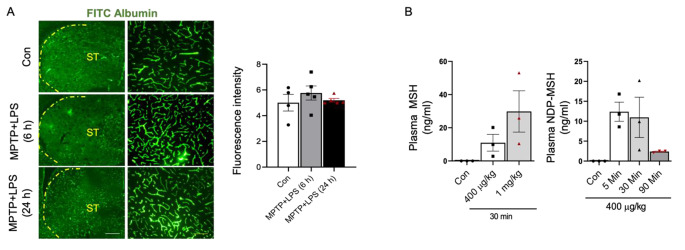
Melanocortin 1 receptor (MC1R) is a key pigmentation gene, and loss-of-function of MC1R variants that produce red hair may be associated with Parkinson's disease (PD). We previously reported compromised dopaminergic neuron survival in Mc1r mutant mice and dopaminergic neuroprotective effects of local injection of a MC1R agonist to the brain or a systemically administered MC1R agonist with appreciable central nervous system (CNS) permeability. Beyond melanocytes and dopaminergic neurons, MC1R is expressed in other peripheral tissues and cell types, including immune cells. The present study investigates the impact of NDP-MSH, a synthetic melanocortin receptor (MCR) agonist that does not cross BBB, on the immune system and the nigrostriatal dopaminergic system in mouse model of PD.
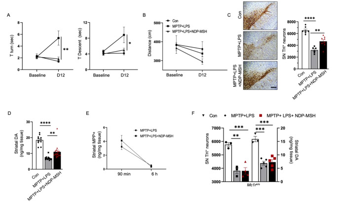
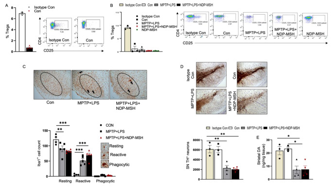
C57BL/6 mice were treated systemically with MPTP.HCl (20 mg/kg) and LPS (1 mg/kg) from day 1 to day 4 and NDP-MSH (400 µg/kg) or vehicle from day 1 to day 12 following which the mice were sacrificed. Peripheral and CNS immune cells were phenotyped and inflammatory markers were measured. The nigrostriatal dopaminergic system was assessed behaviorally, chemically, immunologically, and pathologically. To understand the role of regulatory T cells (Tregs) in this model, CD25 monoclonal antibody was used to deplete CD25 + Tregs.
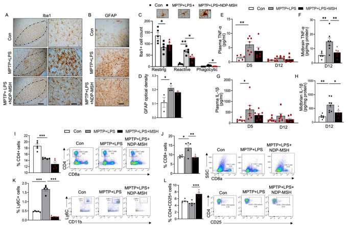
Systemic NDP-MSH administration significantly attenuated striatal dopamine depletion and nigral dopaminergic neuron loss induced by MPTP + LPS. It improved the behavioral outcomes in the pole test. Mc1r mutant mice injected with NDP-MSH in the MPTP and LPS paradigm showed no changes in striatal dopamine levels suggesting that the NDP-MSH acts through the MC1R pathway. Although no NDP-MSH was detected in the brain, peripheral, NDP-MSH attenuated neuroinflammation as observed by diminished microglial activation in the nigral region, along with reduced TNF-α and IL1β levels in the ventral midbrain. Depletion of Tregs was associated with diminished neuroprotective effects of NDP-MSH.
Our study demonstrates that peripherally acting NDP-MSH confers protection on dopaminergic nigrostriatal neurons and reduces hyperactivated microglia. NDP-MSH modulates peripheral immune responses, and Tregs may be involved in the neuroprotective effect of NDP-MSH.
黑素皮质素 1 受体(MC1R)是一个关键的色素基因,产生红发的 MC1R 变体的功能丧失可能与帕金森病(PD)有关。我们之前报道过 Mc1r 突变小鼠中的多巴胺能神经元存活受损,以及局部注射 MC1R 激动剂到大脑或系统给予具有可观中枢神经系统(CNS)通透性的 MC1R 激动剂对多巴胺能神经的保护作用。除黑素细胞和多巴胺能神经元外,MC1R 在其他外周组织和细胞类型中表达,包括免疫细胞。本研究探讨了 NDP-MSH(一种不穿透血脑屏障的合成黑素皮质素受体(MCR)激动剂)对 PD 小鼠模型中免疫系统和黑质纹状体多巴胺能系统的影响。
C57BL/6 小鼠从第 1 天到第 4 天用 MPTP.HCl(20mg/kg)和 LPS(1mg/kg)处理,从第 1 天到第 12 天用 NDP-MSH(400μg/kg)或载体处理,然后处死小鼠。表型分析外周和中枢免疫细胞,测量炎症标志物。黑质纹状体多巴胺能系统通过行为、化学、免疫和病理进行评估。为了了解调节性 T 细胞(Tregs)在该模型中的作用,使用 CD25 单克隆抗体耗尽 CD25+Tregs。
系统给予 NDP-MSH 可显著减轻 MPTP+LPS 诱导的纹状体多巴胺耗竭和黑质多巴胺能神经元丢失。它改善了 pole 测试中的行为结果。在 MPTP 和 LPS 范式中注射 NDP-MSH 的 Mc1r 突变小鼠,纹状体多巴胺水平没有变化,表明 NDP-MSH 通过 MC1R 途径发挥作用。尽管大脑中未检测到 NDP-MSH,但外周 NDP-MSH 减轻了神经炎症,表现为黑质区小胶质细胞激活减少,腹侧中脑 TNF-α和 IL1β水平降低。Tregs 的耗竭与 NDP-MSH 的神经保护作用减弱有关。
我们的研究表明,外周作用的 NDP-MSH 对多巴胺能黑质纹状体神经元具有保护作用,并减少过度激活的小胶质细胞。NDP-MSH 调节外周免疫反应,Tregs 可能参与 NDP-MSH 的神经保护作用。