Department of Cardiology, Affiliated Hospital of Yunnan University, Kunming, 650021, Yunnan, China.
Dali University, Dali, 671003, China.
Eur J Med Res. 2023 Dec 19;28(1):607. doi: 10.1186/s40001-023-01588-4.
Postinfarction cardiac remodeling presents a compensatory mechanism aimed at mitigating congestive heart failure. It is distinguished by progressive dilatation and hypertrophy of the ventricular chambers, fibrotic alterations, and prolonged apoptosis of cardiomyocytes. The primary objective of this study was to assess the effects of icariin on myocardial fibrosis and ventricular remodeling in rats subjected to myocardial infarction (MI).
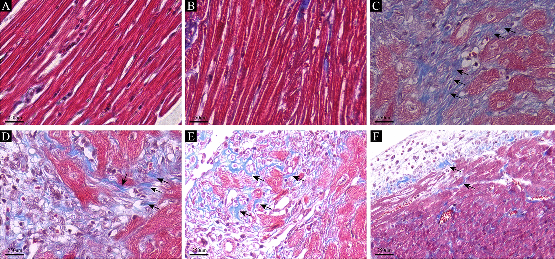
Male Sprague‒Dawley (SD) rats were subjected to randomization and subsequently divided into distinct groups: the control group, the sham group (undergoing sham operation), the MI group (experiencing ligation of the left anterior descending artery), and the icariin group. Within the icariin group, rats were further categorized into three different dose groups based on the administered icariin dosage: the MI30 group (30 mg/kg/day), the MI60 group (60 mg/kg/day), and the MI120 group (120 mg/kg/day). Cardiac function evaluation was carried out using echocardiography. Histological examinations, including hematoxylin and eosin (HE) staining, Masson staining, and immunohistochemistry studies, were conducted 90 days after the occurrence of MI. Additionally, Western blotting was employed to assess TGF-β1, p-Smad2, and p-Smad3 levels.
The administration of icariin revealed a noteworthy enhancement in cardiac function among rats afflicted with left anterior descending coronary artery (LAD) ligation. In comparison to the icariin groups, the MI group exhibited reduced EF and FS, along with elevated LVEDD and LVESD. Furthermore, the cardiac fibrosis levels in the MI group rats exhibited a considerable increase compared to those in the icariin group. Notably, the levels of Collagen I, Collagen III, MMP2, and MMP9 were significantly higher in the MI group than in the icariin group, with evident distinctions. Moreover, the expression levels of TGF-β, IL-13, p-Smad2, and p-Smad3 were notably upregulated in the MI group compared to the icariin group.
In an experimental rat model of MI, the administration of icariin resulted in the amelioration of both cardiac function and remodeling processes, operating through the intricate TGF-β1/Smad signaling pathway.
心肌梗死后心脏重构表现为一种代偿机制,旨在减轻充血性心力衰竭。它的特征是心室腔进行性扩张和肥大、纤维变性改变以及心肌细胞凋亡延长。本研究的主要目的是评估淫羊藿苷对心肌梗死后大鼠心肌纤维化和心室重构的影响。
雄性 Sprague-Dawley(SD)大鼠随机分组,分为对照组、假手术组(仅接受假手术)、心肌梗死组(结扎左前降支)和淫羊藿苷组。在淫羊藿苷组中,根据给予的淫羊藿苷剂量,大鼠进一步分为三个不同剂量组:MI30 组(30mg/kg/天)、MI60 组(60mg/kg/天)和 MI120 组(120mg/kg/天)。使用超声心动图评估心功能。心肌梗死后 90 天进行组织学检查,包括苏木精和伊红(HE)染色、Masson 染色和免疫组织化学研究。此外,还采用 Western blot 检测 TGF-β1、p-Smad2 和 p-Smad3 水平。
淫羊藿苷给药可显著改善左前降支结扎大鼠的心功能。与淫羊藿苷组相比,心肌梗死组 EF 和 FS 降低,LVEDD 和 LVESD 升高。此外,心肌梗死组大鼠的心脏纤维化水平明显高于淫羊藿苷组。值得注意的是,与淫羊藿苷组相比,MI 组大鼠的 Collagen I、Collagen III、MMP2 和 MMP9 水平显著升高,差异明显。此外,与淫羊藿苷组相比,MI 组 TGF-β、IL-1β、p-Smad2 和 p-Smad3 的表达水平明显上调。
在心肌梗死大鼠模型中,淫羊藿苷给药可改善心功能和重构过程,其作用机制可能与 TGF-β1/Smad 信号通路有关。