• 文献检索
  • 文档翻译
  • 深度研究
  • 学术资讯
  • Suppr Zotero 插件Zotero 插件
  • 邀请有礼
  • 套餐&价格
  • 历史记录
应用&插件
Suppr Zotero 插件Zotero 插件浏览器插件Mac 客户端Windows 客户端微信小程序
定价
高级版会员购买积分包购买API积分包
服务
文献检索文档翻译深度研究API 文档MCP 服务
关于我们
关于 Suppr公司介绍联系我们用户协议隐私条款
关注我们

Suppr 超能文献

核心技术专利:CN118964589B侵权必究
粤ICP备2023148730 号-1Suppr @ 2026

文献检索

告别复杂PubMed语法,用中文像聊天一样搜索,搜遍4000万医学文献。AI智能推荐,让科研检索更轻松。

立即免费搜索

文件翻译

保留排版,准确专业,支持PDF/Word/PPT等文件格式,支持 12+语言互译。

免费翻译文档

深度研究

AI帮你快速写综述,25分钟生成高质量综述,智能提取关键信息,辅助科研写作。

立即免费体验

冠状动脉钙化与斑块稳定性:一项光学相干断层扫描研究

Coronary artery calcification and plaque stability: an optical coherence tomography study.

作者信息

Qin Zhifeng, Yu Li, Zhang Yanwen, Xu Qinglu, Li Chao, Zhao Suhong, Xi Xiangwen, Tian Yanan, Wang Zhao, Tian Jinwei, Yu Bo

机构信息

Department of Cardiology, The Second Affiliated Hospital of Harbin Medical University, Harbin, China.

The Key Laboratory of Myocardial Ischemia, Ministry of Education, Harbin, China.

出版信息

Heliyon. 2023 Dec 3;9(12):e23191. doi: 10.1016/j.heliyon.2023.e23191. eCollection 2023 Dec.

DOI:10.1016/j.heliyon.2023.e23191
PMID:38149191
原文链接:https://pmc.ncbi.nlm.nih.gov/articles/PMC10750051/
Abstract

BACKGROUND

Coronary artery calcification (CAC), a surrogate of atherosclerosis, is related to stent underexpansion and adverse cardiac events. However, the effect of CAC on plaque stability is still controversial and the morphological significance of CAC has yet to be elucidated.

METHODS

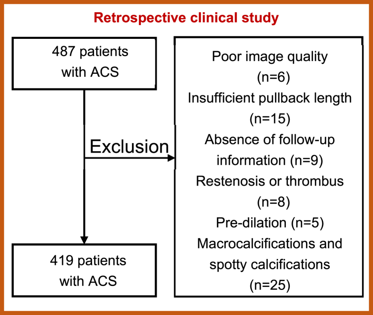
A retrospective series of 419 patients with acute coronary syndrome (ACS) who underwent optical coherence tomography (OCT) were enrolled. Patients were classified into three groups based on the calcification size in culprit plaques and the features of the culprit and non-culprit plaques among these groups were compared. Logistic regression was used to analyze independent risk factors for culprit plaque rupture and the nonlinear relationship between calcification parameters and culprit plaque rupture. Furthermore, we compared the detailed calcification parameters of different kinds of plaques.

RESULTS

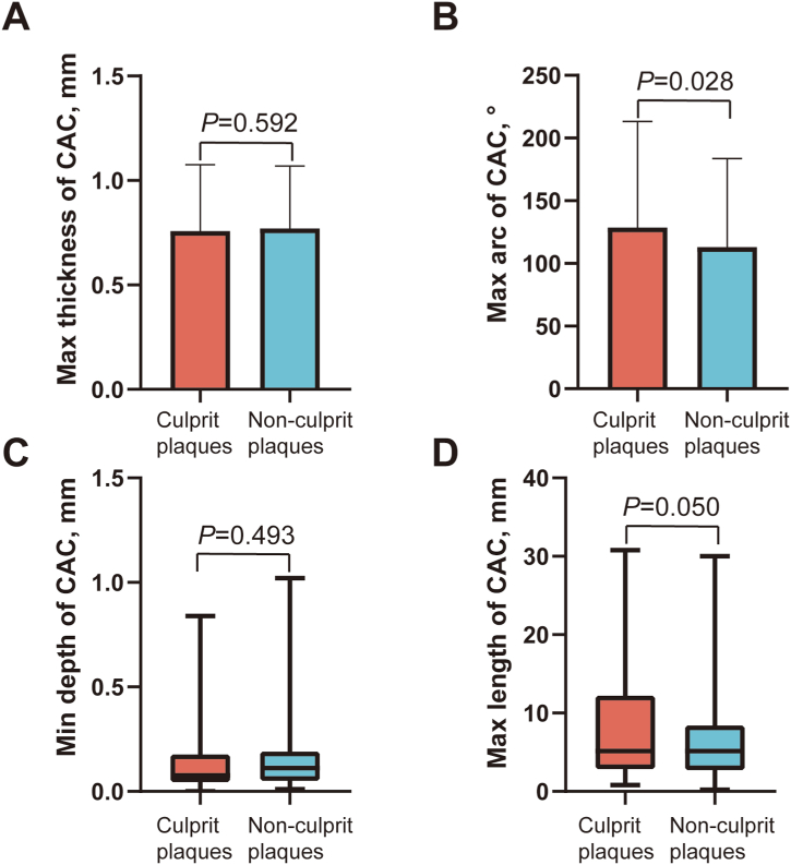
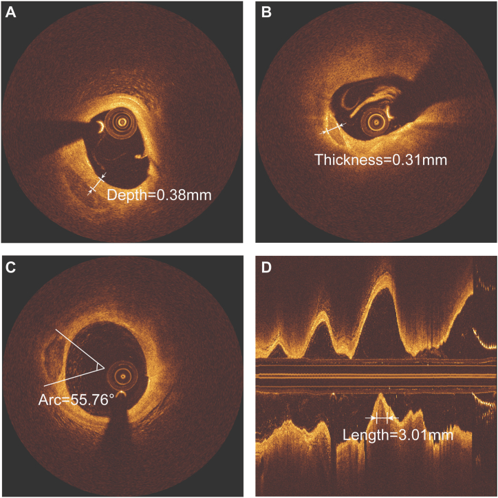
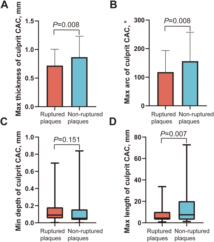
A total of 419 culprit plaques and 364 non-culprit plaques were identified. The incidence of calcification was 53.9 % in culprit plaques and 50.3 % in non-culprit plaques. Compared with culprit plaques without calcification, plaque rupture, macrophages and cholesterol crystals were more frequently observed in the spotty calcification group, and the lipid length was longer; the incidence of macrophages and cholesterol crystals was higher in the macrocalcification group. Calcification tended to be smaller in ruptured plaques than in non-ruptured plaques. Moreover, the arc and length of calcification were greater in culprit plaques than in non-culprit plaques.

CONCLUSIONS

Vulnerable features were more frequently observed in culprit plaques with spotty calcification, whereas the presence of macrocalcification calcifications did not significantly increase plaque vulnerability. Calcification tends to be larger in culprit plaques than in non-culprit plaques.

摘要

背景

冠状动脉钙化(CAC)是动脉粥样硬化的一个替代指标,与支架扩张不全及不良心脏事件相关。然而,CAC对斑块稳定性的影响仍存在争议,其形态学意义也有待阐明。

方法

纳入419例行光学相干断层扫描(OCT)的急性冠状动脉综合征(ACS)患者的回顾性系列研究。根据罪犯斑块中的钙化大小将患者分为三组,并比较这些组中罪犯斑块和非罪犯斑块的特征。采用逻辑回归分析罪犯斑块破裂的独立危险因素以及钙化参数与罪犯斑块破裂之间的非线性关系。此外,我们比较了不同类型斑块的详细钙化参数。

结果

共识别出419个罪犯斑块和364个非罪犯斑块。罪犯斑块中钙化的发生率为53.9%,非罪犯斑块中为50.3%。与无钙化的罪犯斑块相比,斑点状钙化组更常观察到斑块破裂、巨噬细胞和胆固醇结晶,且脂质长度更长;大钙化组中巨噬细胞和胆固醇结晶的发生率更高。破裂斑块中的钙化往往比未破裂斑块中的小。此外,罪犯斑块中钙化的弧度和长度大于非罪犯斑块。

结论

在有斑点状钙化的罪犯斑块中更常观察到易损特征,而大钙化的存在并未显著增加斑块易损性。罪犯斑块中的钙化往往比非罪犯斑块中的大。

https://cdn.ncbi.nlm.nih.gov/pmc/blobs/934a/10750051/935322bab969/gr6.jpg
https://cdn.ncbi.nlm.nih.gov/pmc/blobs/934a/10750051/60d9864d8c63/gr1.jpg
https://cdn.ncbi.nlm.nih.gov/pmc/blobs/934a/10750051/5c478175c2e1/gr2.jpg
https://cdn.ncbi.nlm.nih.gov/pmc/blobs/934a/10750051/5751cfa9e9f7/gr3.jpg
https://cdn.ncbi.nlm.nih.gov/pmc/blobs/934a/10750051/30ca916e807c/gr4.jpg
https://cdn.ncbi.nlm.nih.gov/pmc/blobs/934a/10750051/a3c022c082ee/gr5.jpg
https://cdn.ncbi.nlm.nih.gov/pmc/blobs/934a/10750051/935322bab969/gr6.jpg

相似文献

1
Coronary artery calcification and plaque stability: an optical coherence tomography study.冠状动脉钙化与斑块稳定性:一项光学相干断层扫描研究
Heliyon. 2023 Dec 3;9(12):e23191. doi: 10.1016/j.heliyon.2023.e23191. eCollection 2023 Dec.
2
Cholesterol crystals in non-culprit plaques of STEMI patients: A 3-vessel OCT study.ST段抬高型心肌梗死患者非罪犯斑块中的胆固醇结晶:一项 3 血管 OCT 研究。
Int J Cardiol. 2022 Oct 1;364:162-168. doi: 10.1016/j.ijcard.2022.06.016. Epub 2022 Jun 12.
3
Association between cholesterol crystals and culprit lesion vulnerability in patients with acute coronary syndrome: An optical coherence tomography study.急性冠状动脉综合征患者胆固醇结晶与罪犯病变易损性的关联:一项光学相干断层扫描研究
Atherosclerosis. 2016 Apr;247:111-7. doi: 10.1016/j.atherosclerosis.2016.02.010. Epub 2016 Feb 8.
4
Spotty calcification and plaque vulnerability in vivo: frequency-domain optical coherence tomography analysis.体内斑点状钙化与斑块易损性:频域光学相干断层扫描分析
Cardiovasc Diagn Ther. 2014 Dec;4(6):460-9. doi: 10.3978/j.issn.2223-3652.2014.11.06.
5
Coronary Computed Tomography Angiography Analysis of Calcium Content to Identify Non-culprit Vulnerable Plaques in Patients With Acute Coronary Syndrome.急性冠状动脉综合征患者中通过冠状动脉计算机断层扫描血管造影分析钙含量以识别非罪犯易损斑块
Front Cardiovasc Med. 2022 Apr 15;9:876730. doi: 10.3389/fcvm.2022.876730. eCollection 2022.
6
New insights into spotty calcification and plaque rupture in acute coronary syndrome: an optical coherence tomography study.急性冠状动脉综合征中斑点状钙化和斑块破裂的新见解:一项光学相干断层扫描研究
Heart Vessels. 2016 Dec;31(12):1915-1922. doi: 10.1007/s00380-016-0820-3. Epub 2016 Mar 5.
7
Pancoronary plaque vulnerability in patients with acute coronary syndrome and ruptured culprit plaque: a 3-vessel optical coherence tomography study.急性冠状动脉综合征合并罪犯斑块破裂患者的冠状动脉斑块易损性:三血管光学相干断层成像研究。
Am Heart J. 2014 Jan;167(1):59-67. doi: 10.1016/j.ahj.2013.10.011. Epub 2013 Oct 19.
8
The relationships between cholesterol crystals, NLRP3 inflammasome, and coronary atherosclerotic plaque vulnerability in acute coronary syndrome: An optical coherence tomography study.急性冠状动脉综合征中胆固醇结晶、NLRP3炎性小体与冠状动脉粥样硬化斑块易损性的关系:一项光学相干断层扫描研究
Front Cardiovasc Med. 2022 Oct 25;9:905363. doi: 10.3389/fcvm.2022.905363. eCollection 2022.
9
Differences in the association of total versus local coronary artery calcium with acute coronary syndrome and culprit lesions in patients with acute chest pain: The coronary calcium paradox.在急性胸痛患者中,总冠状动脉钙与局部冠状动脉钙与急性冠状动脉综合征和罪犯病变的关联存在差异:冠状动脉钙悖论。
Atherosclerosis. 2018 Jul;274:251-257. doi: 10.1016/j.atherosclerosis.2018.04.017. Epub 2018 Apr 17.
10
Superficial Calcification With Rotund Shape Is Associated With Carotid Plaque Rupture: An Optical Coherence Tomography Study.圆形浅表钙化与颈动脉斑块破裂相关:一项光学相干断层扫描研究
Front Neurol. 2020 Sep 18;11:563334. doi: 10.3389/fneur.2020.563334. eCollection 2020.

引用本文的文献

1
Study on the association between neovascularization of culprit lesions and plaque rupture in patients with acute myocardial infarction.急性心肌梗死患者罪犯病变新生血管形成与斑块破裂之间的相关性研究。
BMC Cardiovasc Disord. 2025 Aug 2;25(1):567. doi: 10.1186/s12872-025-04995-z.
2
Patient with Vulnerable Coronary Plaque and Treatment with Evolocumab: A Clinical Case.患有易损冠状动脉斑块的患者及依洛尤单抗治疗:一例临床病例
J Clin Med. 2025 Feb 14;14(4):1257. doi: 10.3390/jcm14041257.
3
A Review Paper on Optical Coherence Tomography Evaluation of Coronary Calcification Pattern: Is It Relevant Today?

本文引用的文献

1
Cholesterol crystals in non-culprit plaques of STEMI patients: A 3-vessel OCT study.ST段抬高型心肌梗死患者非罪犯斑块中的胆固醇结晶:一项 3 血管 OCT 研究。
Int J Cardiol. 2022 Oct 1;364:162-168. doi: 10.1016/j.ijcard.2022.06.016. Epub 2022 Jun 12.
2
Optical coherence tomography in coronary atherosclerosis assessment and intervention.光学相干断层成像在冠状动脉粥样硬化评估和干预中的应用。
Nat Rev Cardiol. 2022 Oct;19(10):684-703. doi: 10.1038/s41569-022-00687-9. Epub 2022 Apr 21.
3
Prevalence and extent of coronary artery calcification in the middle-aged and elderly population.
关于冠状动脉钙化模式的光学相干断层扫描评估的综述文章:如今它是否具有相关性?
J Cardiovasc Dev Dis. 2024 Jul 24;11(8):231. doi: 10.3390/jcdd11080231.
中年和老年人的冠状动脉钙化的流行率和程度。
Eur J Prev Cardiol. 2022 Feb 9;28(18):2048-2055. doi: 10.1093/eurjpc/zwab111.
4
Calcification in Atherosclerotic Plaque Vulnerability: Friend or Foe?动脉粥样硬化斑块易损性中的钙化:是友还是敌?
Front Physiol. 2020 Feb 5;11:56. doi: 10.3389/fphys.2020.00056. eCollection 2020.
5
Non-culprit plaque characteristics in acute coronary syndrome patients with raised hemoglobinA1c: an intravascular optical coherence tomography study.糖化血红蛋白升高的急性冠脉综合征患者非罪犯斑块特征:血管内光学相干断层成像研究。
Cardiovasc Diabetol. 2018 Jun 15;17(1):90. doi: 10.1186/s12933-018-0729-5.
6
Predictors for target lesion microcalcifications in patients with stable coronary artery disease: an optical coherence tomography study.稳定型冠状动脉疾病患者靶病变微钙化的预测因素:一项光学相干断层成像研究。
Clin Res Cardiol. 2018 Sep;107(9):763-771. doi: 10.1007/s00392-018-1243-1. Epub 2018 Apr 13.
7
A new optical coherence tomography-based calcium scoring system to predict stent underexpansion.一种基于光学相干断层成像术的新的钙评分系统,用于预测支架扩张不足。
EuroIntervention. 2018 Apr 6;13(18):e2182-e2189. doi: 10.4244/EIJ-D-17-00962.
8
Coronary Artery Calcification and its Progression: What Does it Really Mean?冠状动脉钙化及其进展:它到底意味着什么?
JACC Cardiovasc Imaging. 2018 Jan;11(1):127-142. doi: 10.1016/j.jcmg.2017.10.012.
9
Plaque Calcification During Atherosclerosis Progression and Regression.动脉粥样硬化进展和消退过程中的斑块钙化。
J Atheroscler Thromb. 2018 Apr 1;25(4):294-303. doi: 10.5551/jat.RV17020. Epub 2017 Dec 12.
10
Type 2 diabetes mellitus is associated with a lower fibrous cap thickness but has no impact on calcification morphology: an intracoronary optical coherence tomography study.2 型糖尿病与较薄的纤维帽厚度相关,但对钙化形态无影响:一项冠状动脉光学相干断层成像研究。
Cardiovasc Diabetol. 2017 Dec 1;16(1):152. doi: 10.1186/s12933-017-0635-2.