• 文献检索
  • 文档翻译
  • 深度研究
  • 学术资讯
  • Suppr Zotero 插件Zotero 插件
  • 邀请有礼
  • 套餐&价格
  • 历史记录
应用&插件
Suppr Zotero 插件Zotero 插件浏览器插件Mac 客户端Windows 客户端微信小程序
定价
高级版会员购买积分包购买API积分包
服务
文献检索文档翻译深度研究API 文档MCP 服务
关于我们
关于 Suppr公司介绍联系我们用户协议隐私条款
关注我们

Suppr 超能文献

核心技术专利:CN118964589B侵权必究
粤ICP备2023148730 号-1Suppr @ 2026

文献检索

告别复杂PubMed语法,用中文像聊天一样搜索,搜遍4000万医学文献。AI智能推荐,让科研检索更轻松。

立即免费搜索

文件翻译

保留排版,准确专业,支持PDF/Word/PPT等文件格式,支持 12+语言互译。

免费翻译文档

深度研究

AI帮你快速写综述,25分钟生成高质量综述,智能提取关键信息,辅助科研写作。

立即免费体验

在动物模型中,天然获得的细胞内铜绿假单胞菌在棘阿米巴角膜炎发展中的作用。

The role of naturally acquired intracellular Pseudomonas aeruginosa in the development of Acanthamoeba keratitis in an animal model.

机构信息

School of Optometry and Vision Science, Faculty of Medicine and Health, UNSW, Sydney, Australia.

School of Health and Life Sciences, University of the West of Scotland, Blantyre, Scotland, United Kingdom.

出版信息

PLoS Negl Trop Dis. 2024 Jan 2;18(1):e0011878. doi: 10.1371/journal.pntd.0011878. eCollection 2024 Jan.

DOI:10.1371/journal.pntd.0011878
PMID:38166139
原文链接:https://pmc.ncbi.nlm.nih.gov/articles/PMC10795995/
Abstract

BACKGROUND

Acanthamoeba is an environmental host for various microorganisms. Acanthamoeba is also becoming an increasingly important pathogen as a cause of keratitis. In Acanthamoeba keratitis (AK), coinfections involving pathogenic bacteria have been reported, potentially attributed to the carriage of microbes by Acanthamoeba. This study assessed the presence of intracellular bacteria in Acanthamoeba species recovered from domestic tap water and corneas of two different AK patients and examined the impact of naturally occurring intracellular bacteria within Acanthamoeba on the severity of corneal infections in rats.

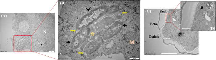
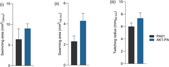
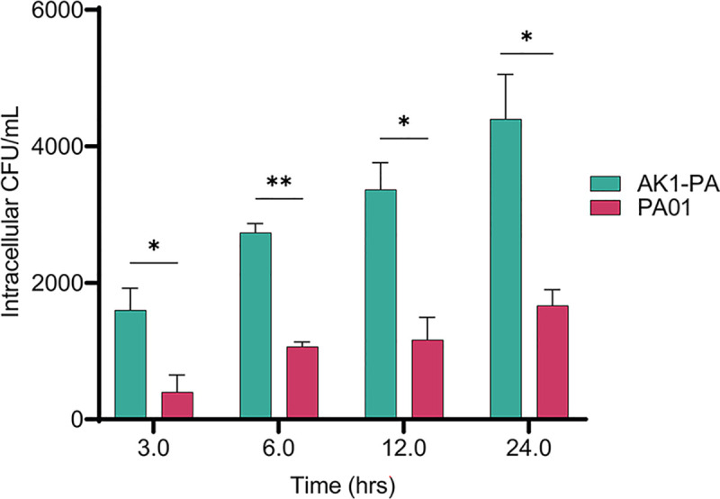
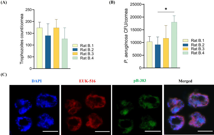
METHODOLOGY/PRINCIPAL FINDINGS: Household water and corneal swabs were collected from AK patients. Acanthamoeba strains and genotypes were confirmed by sequencing. Acanthamoeba isolates were assessed for the presence of intracellular bacteria using sequencing, fluorescence in situ hybridization (FISH), and electron microscopy. The viability of the bacteria in Acanthamoeba was assessed by labelling with alkyne-functionalized D-alanine (alkDala). Primary human macrophages were used to compare the intracellular survival and replication of the endosymbiotic Pseudomonas aeruginosa and a wild type strain. Eyes of rats were challenged intrastromally with Acanthamoeba containing or devoid of P. aeruginosa and evaluated for the clinical response. Domestic water and corneal swabs were positive for Acanthamoeba. Both strains belonged to genotype T4F. One of the Acanthamoeba isolates harboured P. aeruginosa which was seen throughout the Acanthamoeba's cytoplasm. It was metabolically active and could be seen undergoing binary fission. This motile strain was able to replicate in macrophage to a greater degree than strain PAO1 (p<0.05). Inoculation of Acanthamoeba containing the intracellular P. aeruginosa in rats eyes resulted in a severe keratitis with increased neutrophil response. Acanthamoeba alone induced milder keratitis.

CONCLUSIONS/SIGNIFICANCE: Our findings indicate the presence of live intracellular bacteria in Acanthamoeba can increase the severity of acute keratitis in vivo. As P. aeruginosa is a common cause of keratitis, this may indicate the potential for these intracellular bacteria in Acanthamoeba to lead to severe polymicrobial keratitis.

摘要

背景

棘阿米巴是多种微生物的环境宿主。棘阿米巴也正成为一种越来越重要的病原体,可导致角膜炎。在棘阿米巴角膜炎(AK)中,已报道有致病性细菌的合并感染,这可能归因于棘阿米巴携带微生物。本研究评估了从家庭自来水中和两名不同 AK 患者的角膜中分离出的棘阿米巴物种中是否存在细胞内细菌,并研究了自然存在于棘阿米巴内的细胞内细菌对大鼠角膜感染严重程度的影响。

方法/主要发现:从 AK 患者处采集家庭用水和角膜拭子。通过测序确认棘阿米巴株和基因型。使用测序、荧光原位杂交(FISH)和电子显微镜评估棘阿米巴分离株中是否存在细胞内细菌。通过用炔基功能化 D-丙氨酸(alkDala)标记来评估细菌在棘阿米巴内的活力。原代人巨噬细胞用于比较内共生铜绿假单胞菌和野生型菌株的细胞内存活和复制。用含有或不含铜绿假单胞菌的棘阿米巴感染大鼠的基质内,评估临床反应。家庭用水和角膜拭子均为棘阿米巴阳性。两种菌株均属于 T4F 基因型。其中一种棘阿米巴分离株携带铜绿假单胞菌,可在整个棘阿米巴细胞质中看到。它具有代谢活性,并且可以看到正在进行二分分裂。这种运动菌株在巨噬细胞中的复制能力比 PAO1 菌株更强(p<0.05)。在大鼠眼睛中接种含有细胞内铜绿假单胞菌的棘阿米巴会导致严重的角膜炎,伴有中性粒细胞反应增加。单独接种棘阿米巴会引起较轻的角膜炎。

结论/意义:我们的研究结果表明,棘阿米巴内存在活的细胞内细菌会增加体内急性角膜炎的严重程度。由于铜绿假单胞菌是角膜炎的常见病因,这可能表明棘阿米巴内这些细胞内细菌有导致严重多微生物角膜炎的潜力。

https://cdn.ncbi.nlm.nih.gov/pmc/blobs/5a2a/10795995/e876bd873db1/pntd.0011878.g016.jpg
https://cdn.ncbi.nlm.nih.gov/pmc/blobs/5a2a/10795995/865c5b4feaa3/pntd.0011878.g001.jpg
https://cdn.ncbi.nlm.nih.gov/pmc/blobs/5a2a/10795995/884bdd1e42f0/pntd.0011878.g002.jpg
https://cdn.ncbi.nlm.nih.gov/pmc/blobs/5a2a/10795995/f3fb8fc9e36c/pntd.0011878.g003.jpg
https://cdn.ncbi.nlm.nih.gov/pmc/blobs/5a2a/10795995/542bc7817f66/pntd.0011878.g004.jpg
https://cdn.ncbi.nlm.nih.gov/pmc/blobs/5a2a/10795995/d76a5737b2c1/pntd.0011878.g005.jpg
https://cdn.ncbi.nlm.nih.gov/pmc/blobs/5a2a/10795995/944c3352c8cc/pntd.0011878.g006.jpg
https://cdn.ncbi.nlm.nih.gov/pmc/blobs/5a2a/10795995/dde01eecbaa0/pntd.0011878.g007.jpg
https://cdn.ncbi.nlm.nih.gov/pmc/blobs/5a2a/10795995/f1024b79e3b3/pntd.0011878.g008.jpg
https://cdn.ncbi.nlm.nih.gov/pmc/blobs/5a2a/10795995/31c9d8a09aa6/pntd.0011878.g009.jpg
https://cdn.ncbi.nlm.nih.gov/pmc/blobs/5a2a/10795995/e9ca979796b5/pntd.0011878.g010.jpg
https://cdn.ncbi.nlm.nih.gov/pmc/blobs/5a2a/10795995/26391edc1e7f/pntd.0011878.g011.jpg
https://cdn.ncbi.nlm.nih.gov/pmc/blobs/5a2a/10795995/be4ab21aa5f1/pntd.0011878.g012.jpg
https://cdn.ncbi.nlm.nih.gov/pmc/blobs/5a2a/10795995/e7fa6fc43d93/pntd.0011878.g013.jpg
https://cdn.ncbi.nlm.nih.gov/pmc/blobs/5a2a/10795995/cd41cf8b776e/pntd.0011878.g014.jpg
https://cdn.ncbi.nlm.nih.gov/pmc/blobs/5a2a/10795995/6b2f35658e77/pntd.0011878.g015.jpg
https://cdn.ncbi.nlm.nih.gov/pmc/blobs/5a2a/10795995/e876bd873db1/pntd.0011878.g016.jpg

相似文献

1
The role of naturally acquired intracellular Pseudomonas aeruginosa in the development of Acanthamoeba keratitis in an animal model.在动物模型中,天然获得的细胞内铜绿假单胞菌在棘阿米巴角膜炎发展中的作用。
PLoS Negl Trop Dis. 2024 Jan 2;18(1):e0011878. doi: 10.1371/journal.pntd.0011878. eCollection 2024 Jan.
2
Assessment of genotypes, endosymbionts and clinical characteristics of Acanthamoeba recovered from ocular infection.从眼部感染中分离的棘阿米巴的基因型、内共生体和临床特征评估。
BMC Infect Dis. 2022 Sep 29;22(1):757. doi: 10.1186/s12879-022-07741-4.
3
Number of Bacteria and Time of Coincubation With Bacteria Required for the Development of Acanthamoeba Keratitis.棘阿米巴角膜炎发生所需的细菌数量及与细菌共培养的时间
Cornea. 2017 Mar;36(3):353-357. doi: 10.1097/ICO.0000000000001129.
4
The role of domestic tap water on Acanthamoeba keratitis in non-contact lens wearers and validation of laboratory methods.家用自来水在非隐形眼镜佩戴者棘阿米巴角膜炎中的作用及实验室方法的验证
Parasitol Res. 2015 Sep;114(9):3283-9. doi: 10.1007/s00436-015-4549-1. Epub 2015 May 29.
5
Corticosteroid eye drop instillation aggravates the development of Acanthamoeba keratitis in rabbit corneas inoculated with Acanthamoeba and bacteria.皮质类固醇滴眼剂的滴注加重了接种棘阿米巴和细菌的兔角膜中棘阿米巴角膜炎的发展。
Sci Rep. 2019 Sep 6;9(1):12821. doi: 10.1038/s41598-019-49128-7.
6
Detection of bacterial endosymbionts in clinical acanthamoeba isolates.临床棘阿米巴分离株中细菌内共生体的检测。
Ophthalmology. 2010 Mar;117(3):445-52, 452.e1-3. doi: 10.1016/j.ophtha.2009.08.033. Epub 2010 Jan 19.
7
Investigation of the Role of Bacteria in the Development of Acanthamoeba Keratitis.细菌在棘阿米巴角膜炎发病过程中的作用研究。
Cornea. 2015 Oct;34(10):1308-15. doi: 10.1097/ICO.0000000000000541.
8
Acanthamoeba keratitis in noncompliant soft contact lenses users: Genotyping and risk factors, a study from Cairo, Egypt.埃及开罗一项研究:不合规软性隐形眼镜佩戴者棘阿米巴角膜炎:基因分型和危险因素。
J Infect Public Health. 2018 May-Jun;11(3):377-383. doi: 10.1016/j.jiph.2017.09.013. Epub 2017 Sep 28.
9
Drug-resistant severe Acanthamoeba keratitis caused by rare T5 Acanthamoeba genotype.耐药性严重棘阿米巴角膜炎由罕见的 T5 棘阿米巴基因型引起。
Eye Contact Lens. 2010 May;36(3):183-4. doi: 10.1097/icl.0b013e3181da2350.
10
Genotypic, physiological, and biochemical characterization of potentially pathogenic Acanthamoeba isolated from the environment in Cairo, Egypt.从埃及开罗环境中分离出的潜在致病性棘阿米巴的基因型、生理学和生化特征。
Parasitol Res. 2016 May;115(5):1871-81. doi: 10.1007/s00436-016-4927-3. Epub 2016 Feb 3.

引用本文的文献

1
The hidden dancers in water: the symbiotic mystery of and free-living amoebae.水中的隐藏舞者:纤毛虫与自由生活变形虫的共生之谜
Front Microbiol. 2025 Aug 8;16:1634806. doi: 10.3389/fmicb.2025.1634806. eCollection 2025.
2
The pathogenesis, risk factors, diagnosis and treatment of Acanthamoeba keratitis.棘阿米巴角膜炎的发病机制、危险因素、诊断与治疗
Front Med (Lausanne). 2025 Jul 24;12:1559224. doi: 10.3389/fmed.2025.1559224. eCollection 2025.
3
Achromobacter in the Conjunctival Sac Microbiota: Potential Association With Acanthamoeba Keratitis Related to Orthokeratology Lenses.

本文引用的文献

1
Experimental Induction of Acute Acanthamoeba castellanii Keratitis in Cats.实验诱导猫急性棘阿米巴角膜炎。
Transl Vis Sci Technol. 2023 Aug 1;12(8):10. doi: 10.1167/tvst.12.8.10.
2
Identification and quantification of Acanthamoeba spp. within seawater at four coastal lagoons on the east coast of Australia.澳大利亚东海岸四个沿海泻湖海水中棘阿米巴属物种的鉴定与定量分析。
Sci Total Environ. 2023 Nov 25;901:165862. doi: 10.1016/j.scitotenv.2023.165862. Epub 2023 Aug 2.
3
A 5-Year Review of Coinfections in Acanthamoeba keratitis From South India.
结膜囊微生物群中的无色杆菌:与角膜塑形镜相关的棘阿米巴角膜炎的潜在关联。
Invest Ophthalmol Vis Sci. 2025 Jul 1;66(9):71. doi: 10.1167/iovs.66.9.71.
4
Clinical presentation, genotypic diversity, and intracellular bacteria in keratitis patients treated at a referral eye hospital in Sydney, Australia.澳大利亚悉尼一家转诊眼科医院治疗的角膜炎患者的临床表现、基因型多样性和细胞内细菌
IJID Reg. 2025 Jun 15;16:100690. doi: 10.1016/j.ijregi.2025.100690. eCollection 2025 Sep.
5
First report on the infection of and in the endangered Chinese Milu deer () in China.中国濒危麋鹿()感染和的首次报告。 (注:原文中“ and ”处信息缺失,不太完整准确)
One Health. 2025 Jun 13;21:101112. doi: 10.1016/j.onehlt.2025.101112. eCollection 2025 Dec.
6
: disease-causing pathogen or opportunistic bystander?致病病原体还是机会性旁观者?
J Med Microbiol. 2025 Mar;74(3). doi: 10.1099/jmm.0.001953.
7
Harnessing Non-Antibiotic Strategies to Counter Multidrug-Resistant Clinical Pathogens with Special Reference to Antimicrobial Peptides and Their Coatings.利用非抗生素策略对抗多重耐药临床病原体,特别提及抗菌肽及其涂层
Antibiotics (Basel). 2025 Jan 9;14(1):57. doi: 10.3390/antibiotics14010057.
8
Isolation of Acanthamoeba Species and Bacterial Symbiont Variability in Puna Salt Plains, Argentina.阿根廷普纳盐沼中棘阿米巴属物种的分离及细菌共生体变异性研究
Environ Microbiol Rep. 2025 Feb;17(1):e70059. doi: 10.1111/1758-2229.70059.
9
Unravelling mechanisms of bacterial recognition by Acanthamoeba: insights into microbial ecology and immune responses.揭示棘阿米巴对细菌的识别机制:对微生物生态学和免疫反应的见解
Front Microbiol. 2024 Aug 23;15:1405133. doi: 10.3389/fmicb.2024.1405133. eCollection 2024.
10
First Report of Genotype T4 from the Newly Formed Tajogaite Volcano Tephra (La Palma, Canary Islands).来自新形成的塔约加特火山火山灰(加那利群岛拉帕尔马岛)的T4基因型首次报告。
Pathogens. 2024 Jul 27;13(8):626. doi: 10.3390/pathogens13080626.
印度南部棘阿米巴角膜炎合并感染的 5 年回顾
Eye Contact Lens. 2023 Aug 1;49(8):334-338. doi: 10.1097/ICL.0000000000001005. Epub 2023 May 24.
4
Establishing an experimental Pseudomonas aeruginosa keratitis model in mice - Challenges and solutions.建立小鼠铜绿假单胞菌角膜炎模型 - 挑战与解决方案。
Ann Anat. 2023 Aug;249:152099. doi: 10.1016/j.aanat.2023.152099. Epub 2023 Apr 25.
5
The global epidemiology and clinical diagnosis of Acanthamoeba keratitis.棘阿米巴角膜炎的全球流行病学和临床诊断。
J Infect Public Health. 2023 Jun;16(6):841-852. doi: 10.1016/j.jiph.2023.03.020. Epub 2023 Mar 23.
6
Assessment of genotypes, endosymbionts and clinical characteristics of Acanthamoeba recovered from ocular infection.从眼部感染中分离的棘阿米巴的基因型、内共生体和临床特征评估。
BMC Infect Dis. 2022 Sep 29;22(1):757. doi: 10.1186/s12879-022-07741-4.
7
Acanthamoeba, an environmental phagocyte enhancing survival and transmission of human pathogens.棘阿米巴,一种环境吞噬细胞,增强了人类病原体的存活和传播。
Trends Parasitol. 2022 Nov;38(11):975-990. doi: 10.1016/j.pt.2022.08.007. Epub 2022 Sep 12.
8
Acanthamoeba keratitis: an increasingly common infectious disease of the cornea.棘阿米巴角膜炎:一种日益常见的角膜感染性疾病。
Lancet Microbe. 2021 Aug;2(8):e345-e346. doi: 10.1016/S2666-5247(21)00093-8. Epub 2021 May 25.
9
Advances in the management of Acanthamoeba keratitis: A review of the literature and synthesized algorithmic approach.棘阿米巴角膜炎的治疗进展:文献综述与综合算法方法。
Ocul Surf. 2022 Jul;25:26-36. doi: 10.1016/j.jtos.2022.04.003. Epub 2022 Apr 22.
10
Case Series: Unusual Presentation of Acanthamoeba Coinfection in the Cornea.病例系列:角膜中棘阿米巴共感染的不寻常表现。
Optom Vis Sci. 2022 Jul 1;99(7):605-611. doi: 10.1097/OPX.0000000000001906. Epub 2022 Apr 13.